Glucagon antibody was prepared from whole rabbit serum produced by repeated immunizations with a synthetic peptide corresponding to an internal portion of human Glucagon.
0.02 M Potassium Phosphate, 0.15 M Sodium Chloride, pH 7.2
Formulierung:
Liquid (sterile filtered)
Target-Kategorie:
Human
Antibody Type:
Primary Antibody
Application Verdünnung:
ELISA: 1:24,800-1:44,800, IHC: 1:100, IF Microscopy: 5-15µg/mL, WB: User Optimized
Anwendungsbeschreibung:
Anti-Glucagon is tested in ELISA, IF, and Western Blot. Although not tested, this antibody is suitable in immunohistochemistry. Expect a band approximately ~20.9 kDa corresponding to the appropriate cell lysate or extract. Specific conditions for reactiv
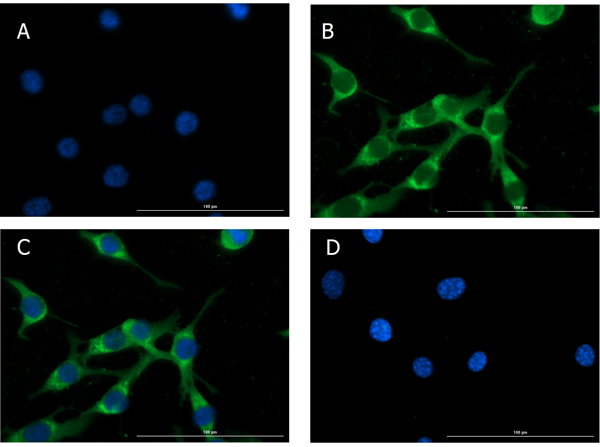
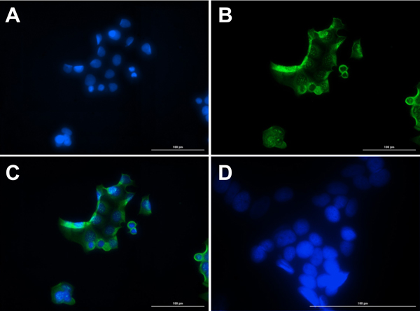
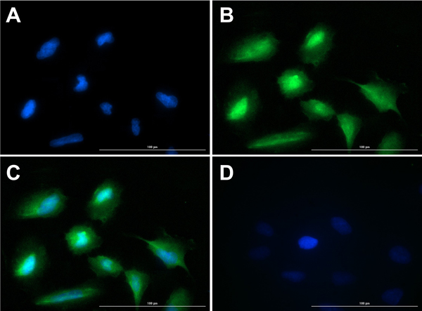
Immunofluorescence of Rabbit Anti-Glucagon Antibody. Cell Line: NIH/3T3 cells. Fixative: 4% PFA. Permeabilization: Triton X-100. Primary Antibody: Anti-Glucagon at 15µg/mL overnight at 2-8C. Secondary Antibody: Goat Anti-Rabbit IgG DyLight(TM)488 (p/n 611-141-122) at 5µL/mL for 1hr at RT. Nuclear Counterstain: DAPI. Staining: (A). DAPI. (B). Anti-Glucagon + DyLight(TM)488 secondary. (C). Merge A+B. (D). secondary only. Localization expected: Cytoplasm.
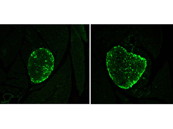
Immunohistochemistry results using Rabbit Anti-Glucagon Antibody. Tissue: alpha cells in CD1 mouse pancreatic islets. Fixation: 4% paraformaldehyde. Antigen Retrieval: 10mM Sodium Citrate buffer for 10 mins at 95-100C. Blocking: PBS, 1% ovalbumin, 0.3% Triton X-100. Primary Antibody: Anti-Glucagon at 1:100 overnight at RT. Secondary Antibody: Anti-Rabbit Alexa Fluor 488 at 1:500 for 1hr at RT. Original magnification 20x. Independently Validated by antibodies-online GmbH (ABIN7448121). Courtesy of Prof. Merighi, University of Turin.
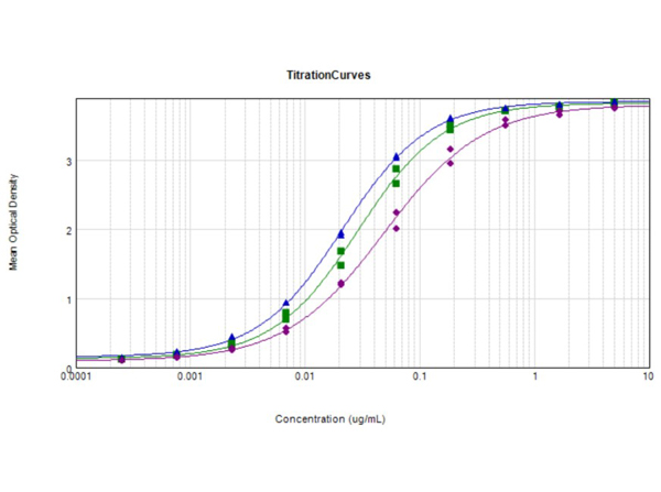
ELISA Results of Rabbit Anti-Glucagon Antibody. Each well was coated with 1µg of conjugate. The starting concentration of antibody in the dilution series was 5 µg/ml. The titer is 1:34,800 Glucagon - Free peptide [Green Line], 1:47200 Glucagon Standard - BSA conjugated [Blue Line], and 1:20,500 Glucagon - BSA conjugated [Purple Line]. Each point on the Y-axis represents a 3-fold dilution. 3% Fish Gel (p/n MB-066-0100), HRP conjugated Goat anti-Rabbit IgG (H&L) (p/n 611-1302), and TMB substrate (p/n TMB-1000) were used for detection.
Immunofluorescence of Rabbit Anti-Glucagon Antibody. Cell Line: NIH/3T3 cells. Fixative: 100% Methanol. Permeabilization: Triton X-100. Primary Antibody: Anti-Glucagon at 15µg/mL overnight at 2-8C. Secondary Antibody: Goat Anti-Rabbit IgG DyLight(TM)488 (p/n 611-141-122) at 5µL/mL for 1hr at RT. Nuclear Counterstain: DAPI. Staining: (A). DAPI. (B). Anti-Glucagon + DyLight(TM)488 secondary. (C). Merge A+B. (D). secondary only. Localization expected: Cytoplasm.
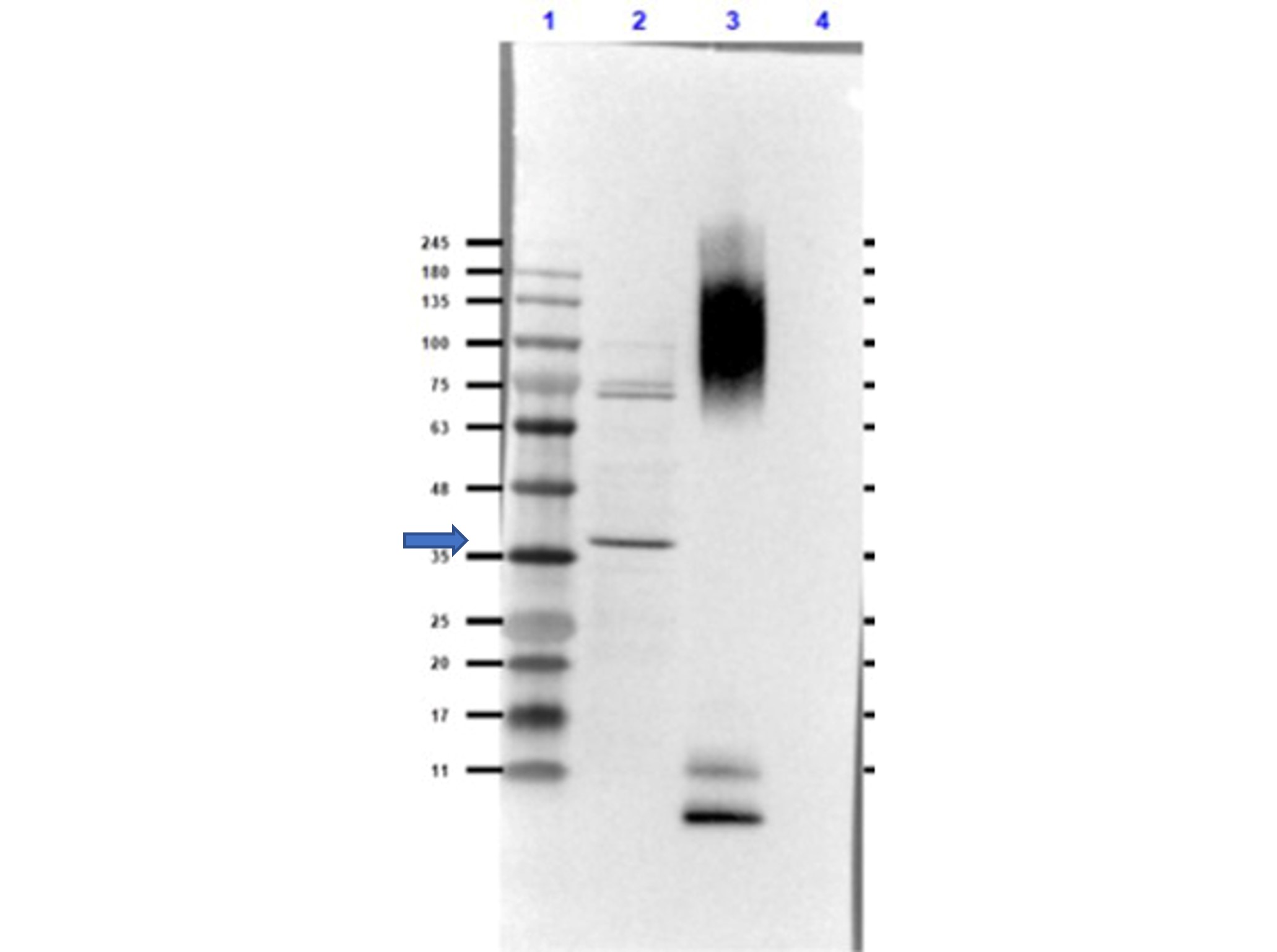
Western Blot of Rabbit Anti-Glucagon Antibody. Lane 1: Opal Prestained Molecular Weight (p/n MB-210-0500). Lane 2: COS-7 Lysate - reduced (20µg). Lane 3: BSA Conjugated Glucagon peptide - reduced (0.02µg).Lane 4: Insulin - reduced (0.05µg). Primary Antibody: Anti-Glucagon [Rabbit] Antibody at 1.0µg/mL overnight at 2-8C. Secondary Antibody: Goat Anti-Rabbit IgG (MX10) Peroxidase conjugated at 1:70,000 for 30mins at RT. Block: Blocking Buffer for Fluorescent Western Blotting (p/n MB-070) for 1hr at RT.Expected MW: ~21kDa. Observed MW: endogenous detection in COS-7 Lysate at ~35kDa. Glucagon peptide is detected at the MW of BSA. No cross-reactivity with insulin is observed.Exposure: 25 sec.
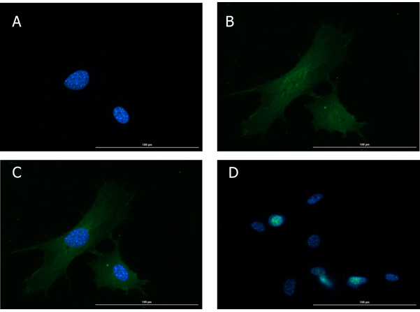
Immunofluorescence of Rabbit Anti-Glucagon Antibody. Cell Line: MCF7 cells. Fixative: 100% Methanol. Permeabilization: 0.3% Triton X-100. Primary Antibody: Anti-Glucagon at 15µg/mL overnight at 2-8C. Secondary Antibody: Goat Anti-Rabbit IgG DyLight(TM)488 (p/n 611-141-122) at 5µL/mL for 1hr at RT. Nuclear Counterstain: DAPI. Staining: (A). DAPI. (B). Anti-Glucagon + DyLight(TM)488 secondary. (C). Merge A+B. (D). secondary only. Localization expected: Cytoplasm.
Immunofluorescence of Rabbit Anti-Glucagon Antibody. Cell Line: U20S cells. Fixative: 4% PFA. Permeabilization: 0.3% Triton X-100. Primary Antibody: Anti-Glucagon at 15µg/mL overnight at 2-8C. Secondary Antibody: Goat Anti-Rabbit IgG DyLight(TM)488 (p/n 611-141-122) at 5µL/mL for 1hr at RT. Nuclear Counterstain: DAPI. Staining: (A). DAPI. (B). Anti-Glucagon + DyLight(TM)488 secondary. (C). Merge A+B. (D). secondary only. Localization expected: Cytoplasm.
* Mehrwertsteuer und Versandkosten nicht enthalten. Irrtümer und Preisänderungen vorbehalten