Anti-ACE2 antibody was prepared from whole rabbit serum produced by repeated immunizations with a synthetic peptide corresponding to amino acids near the internal region of human ACE2.
Konjugation:
Unconjugated
Alternative Synonym:
ACE2 Antibody, ACEH, Angiotensin-converting enzyme 2, ACE-related carboxypeptidase, ACEH
Anti-ACE2 Antibody has been tested for use in ELISA, Immunofluorescence, Immunohistochemistry and Western Blotting. WB has been validated in human mouse, and rat samples, IHC and IF has been validated in human samples. Expect a band at ~93 kDa in size co
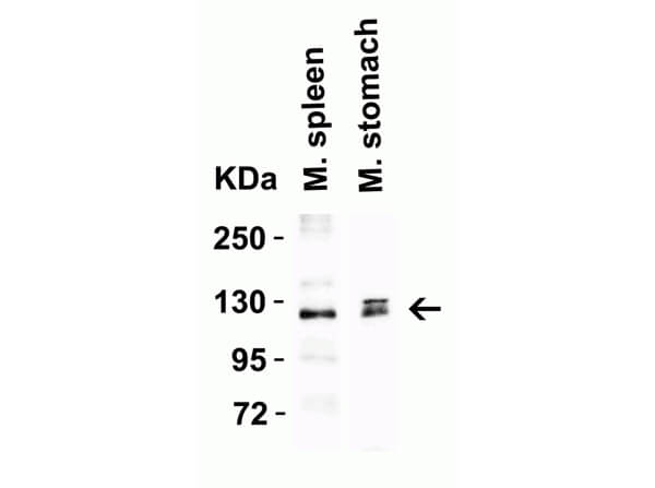
Western Blot of Anti-ACE2 Antibody. Lane 1: Mouse Spleen Lysate. Lane 2: Mouse Stomach Lysate. Load: 15 µg of lysates per lane. Primary Antibody: Anti-ACE2 2µg/mL for 1hr at RT in 5% BLOTTO/TBST. Secondary: Goat anti-rabbit IgG HRP conjugated at 1:10,000. Predicted MW: ~93kDa. Observed: ~125kDa.
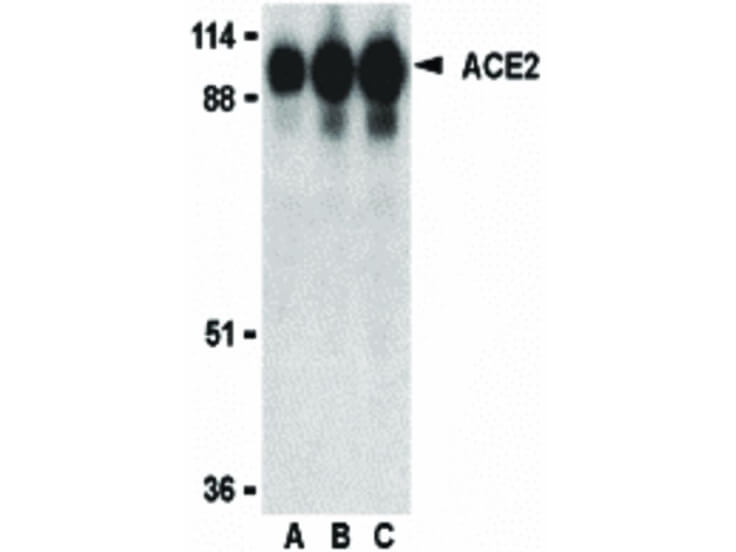
Western Blot of ACE2 antibody. Lane A: human kidney lysate at 0.5 µg/ml. Lane B: human kidney lysate at 1 µg/ml. Lane C: human kidney lysate at 2 µg/ml. Load: 35 µg per lane. Primary antibody: ACE2 antibody at designated concentrations for overnight at 4C. Secondary antibody: Peroxidase rabbit secondary antibody at 1:10,000 for 45 min at RT. Block: 5% BLOTTO overnight at 4C. Predicted MW: 93 kDa. Observed MW: 95 kDa for ACE2. Other band(s): ACE2 splice variants and isoforms.
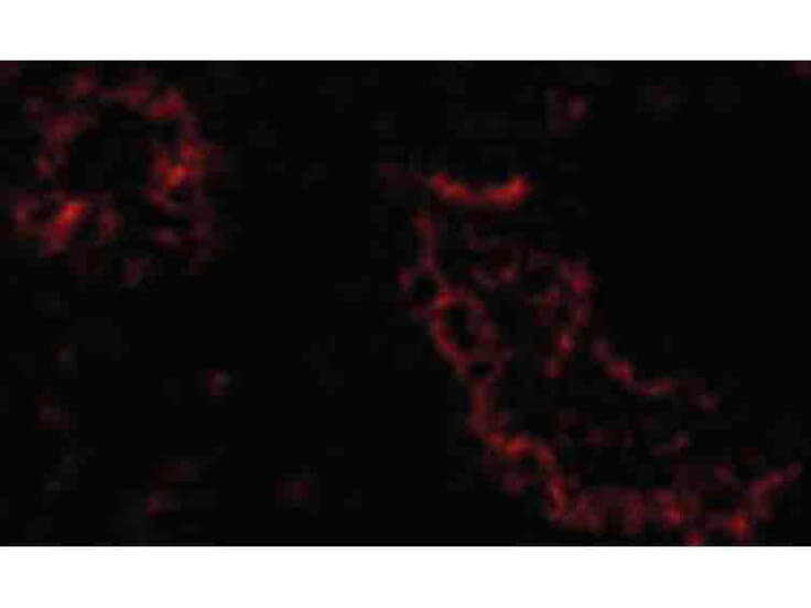
Immunofluorescence Microscopy of ACE2 antibody. Cell Type: human kidney cells. Fixation: 0.5% PFA. Antigen retrieval: not required. Primary antibody: ACE2 antibody at 20 µg/mL for 1 h at RT. Secondary antibody: Fluorescein rabbit secondary antibody at 1:10,000 for 45 min at RT. Localization: ACE2 is located in the cell membrane and the cytoplasm. Staining: ACE2 as red fluorescent signal.
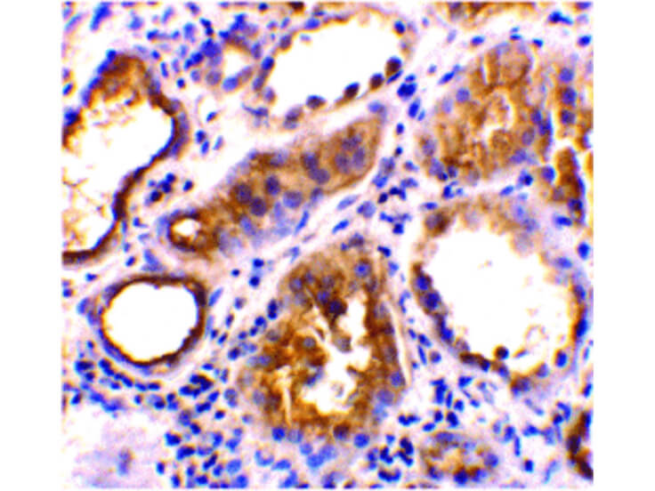
Immunohistochemistry of ACE2 antibody. Tissue: human kidney tissue. Fixation: formalin fixed paraffin embedded. Antigen retrieval: not required. Primary antibody: ACE2 antibody at 2 µg/mL for 1 h at RT. Secondary antibody: Peroxidase rabbit secondary antibody at 1:10,000 for 45 min at RT. Localization: ACE2 is located in the cell membrane and the cytoplasm. Staining: ACE2 as precipitated red signal.
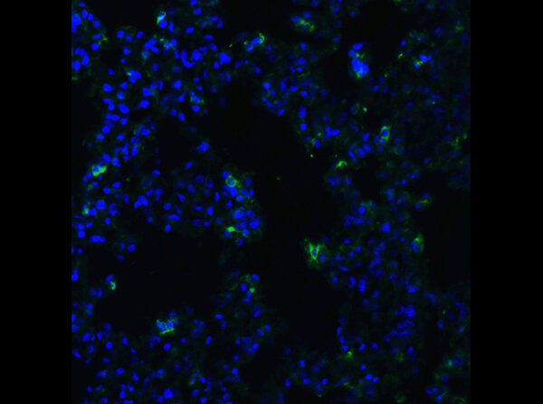
Immunofluorescence of Rb Anti-ACE2 Antibody. Tissue: Human Lung Tissue. Fixation: 4% PFA. Primary Antibody: Anti-ACE2 at 20µg/mL. Secondary Antibody: Goat anti-rabbit IgG secondary antibody at 1:500 dilution (green) and DAPI counterstain (blue).
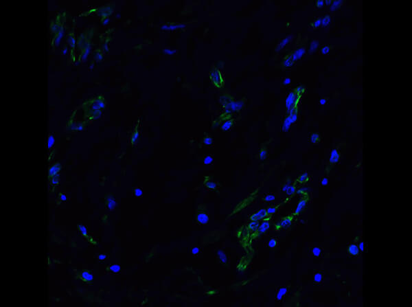
Immunofluorescence of Anti-ACE2 Antibody. Tissue: Rat Lung Tissue. Fixation: 4% PFA. Primary Antibody: Anti-ACE2 at 20µg/mL. Secondary Antibody: Goat anti-rabbit IgG secondary antibody at 1:500 dilution (green) and DAPI counterstain (blue).
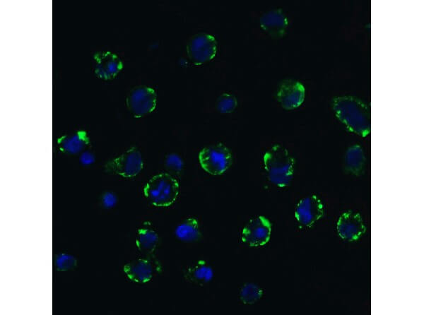
Immunofluorescence of Rabbit Anti-ACE2 Antibody. Tissue: Caco2 cells. Fixation: 4% PFA. Primary antibody: Anti-ACE2 at 20µg/mL. Secondary Antibody: Goat Anti-rabbit IgG secondary antibody at 1:500 dilution (green) and DAPI counterstain (blue). Image showing membrane staining on Caco2 cells.
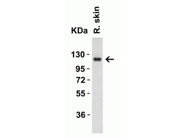
Western Blot of Rabbit Anti-ACE2 Antibody. Lane 1: Rat Skin Lysate. Load: 15 µg of lysates per lane. Primary Antibody: Anti-ACE2 at 2µg/mL for 1h at RT in 5% BLOTTO/TBST. Secondary: Goat anti-rabbit IgG HRP conjugate at 1:10000 dilution. Predicted MW: ~93kDa. Observed MW: ~125kDa.
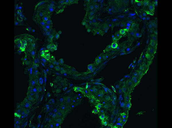
Immunofluorescence of Rabbit Anti-ACE2 Antibody. Tissue: Human Testis Tissue. Fixation: 4% PFA. Primary antibody: Anti-ACE2 at 20µg/mL. Secondary Antibody: Goat anti-rabbit IgG secondary antibody at 1:500 dilution (green) and DAPI counterstain (blue).
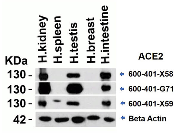
Western Blot of different Rabbit anti-ACE2 antibodies. Lane 1: Human Kidney Lysate. Lane 2: Human Spleen Lysate. Lane 3: Human Testis Lysate. Lane 4: Human Breast Lysate. Lane 5: Human Intestine Lysate. Load: 15 µg per lane. Primary antibody: ACE2 antibody (600-401-X58, 600-401-G71, 600-401-X59) at 2µg/mL and Beta Actin at 1µg/mL for 1hr at RT. Secondary antibody: Goat anti-Rabbit secondary HRP antibody. Block: 5% BLOTTO/TBST. Predicted MW: ~93kDa. Observed: ~130kDa.
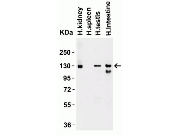
Western Blot of Rabbit anti-ACE2 Antibody. Lane 1: Human Kidney Lysate. Lane 2: Human Spleen Lysate. Lane 3: Human Testis Lysate. Lane 4: Human Intestine Lysate. Loading: 15µg of lysates per lane. Primary Antibody: Anti-ACE2 at 2µg/mL for 1hr at RT in 5% BLOTTO/TBST. Secondary: Goat anti-rabbit IgG HRP conjugate at 1:10000 dilution. Predicted MW: ~93kDa. Observed MW: ~130 kDa.
* Mehrwertsteuer und Versandkosten nicht enthalten. Irrtümer und Preisänderungen vorbehalten