Anti-ACE2 antibody was prepared from whole rabbit serum produced by repeated immunizations with a synthetic peptide corresponding to amino acids near the N terminus of human ACE2.
Konjugation:
Unconjugated
Alternative Synonym:
ACE2 Antibody, ACEH, Angiotensin-converting enzyme 2, ACE-related carboxypeptidase, ACEH
0.01 M Sodium Phosphate, 0.25 M Sodium Chloride, pH 7.2
Formulierung:
Liquid (sterile filtered)
Target-Kategorie:
Human
Antibody Type:
Primary Antibody
Application Verdünnung:
ELISA: 1:10,000, IHC: 2µg/mL, IF Microscopy: 10µg/mL, WB: 1 µg/mL
Anwendungsbeschreibung:
Anti-ACE2 Antibody has been tested for use in ELISA, Immunofluorescence, Immunohistochemistry and Western Blotting. WB validated in human, mouse, and rat samples. IHC and IF validated in human samples. Expect a band at ~93 kDa in size corresponding to AC
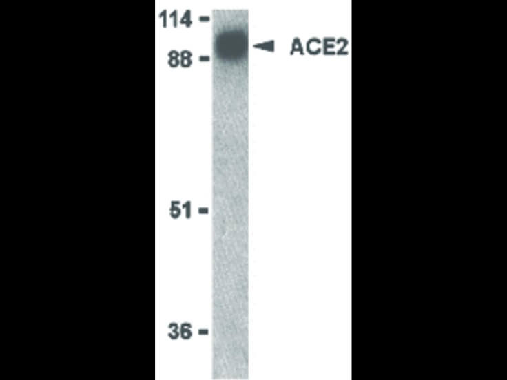
Western Blot of ACE2 antibody. Load: human kidney lysate 35 µg per lane. Primary antibody: ACE2 antibody at 1µg/ml for overnight at 4C. Secondary antibody: Peroxidase rabbit secondary antibody at 1:10,000 for 45 min at RT. Block: 5% BLOTTO overnight at 4C. Predicted/Observed size: 92 kDa, 95 kDa for ACE2. Other band(s): ACE2 splice variants and isoforms.
Immunofluorescence of Rabbit Anti-ACE2 Antibody. Tissue: Human Lung Tissue. Fixation: 4% PFA. Primary Antibody: Anti-ACE2 at 20µg/mL. Secondary Antibody: Goat anti-rabbit IgG secondary antibody at 1:500 dilution (green) and DAPI counterstain (blue).
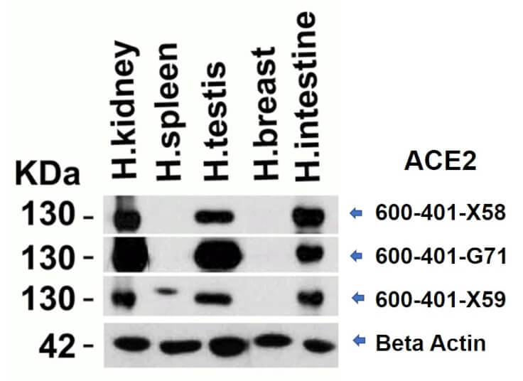
Western Blot of Rabbit Anti-ACE2 Antibody. Lane 1: Human Kidney Lysate. Lane 2: Human Spleen Lysate. Lane 3: Human Testis Lysate. Lane 4: Human Breast Lysate. Lane 5: Human Intestine Lysate. Load: 15µg of lysates per lane. Primary Antibody: ACE2 (600-401-X58, 600-401-G71, 600-401-X59) at 2µg/mL and Beta Actin at 1µg/mL for 1hr at RT in 5% BLOTTO/TBST. Secondary Antibody: Goat anti-rabbit IgG HRP conjugate at 1:10000 dilution. Predicted MW: ~93kDa. Observed MW: ~130kDa.
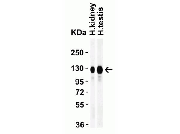
Western Blot of Rabbit Anti-ACE2 Antibody. Lane 1: Human Kidney Lysate. Lane 2: Human Testis Lysate. Load: 15µg of lysates per lane. Primary Antibody: Anti-ACE2 at 2µg/mL for 1hr at RT in 5% BLOTTO/TBST. Secondary Antibody: Goat anti-rabbit IgG HRP conjugate at 1:10000 dilution. Predicted MW: ~93kDa.
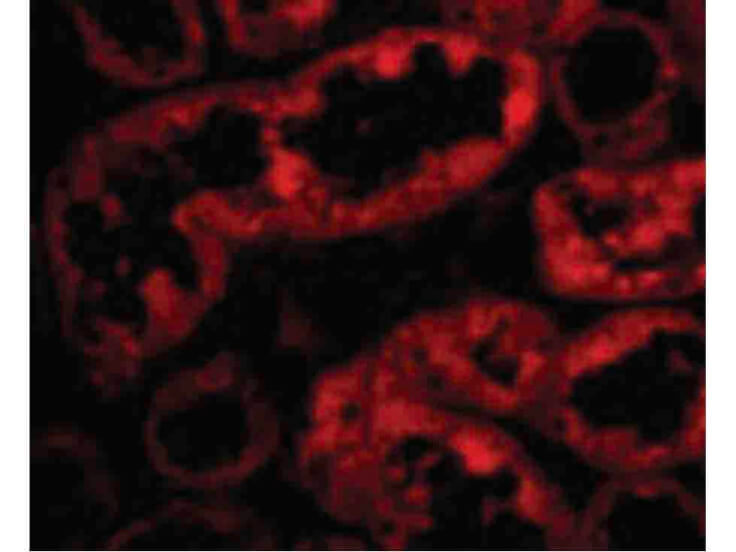
Immunofluorescence Microscopy of ACE2 antibody. Cell Type: human kidney cells. Fixation: 0.5% PFA. Antigen retrieval: not required. Primary antibody: ACE2 antibody at 10 µg/mL for 1 h at RT. Secondary antibody: Fluorescein rabbit secondary antibody at 1:10,000 for 45 min at RT. Localization: ACE2 is located in the cell membrane and cytoplasm. Staining: ACE2 as red fluorescent signal.
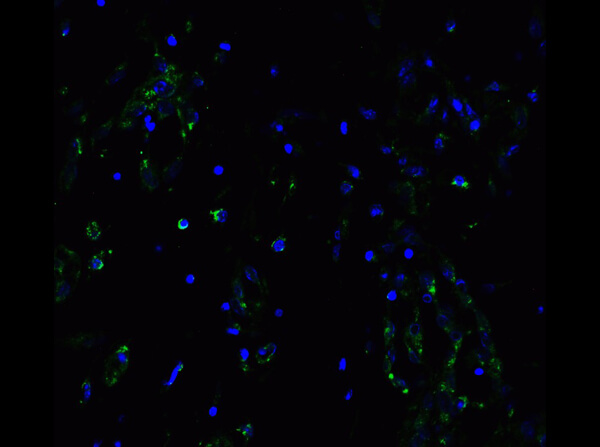
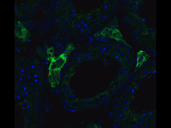
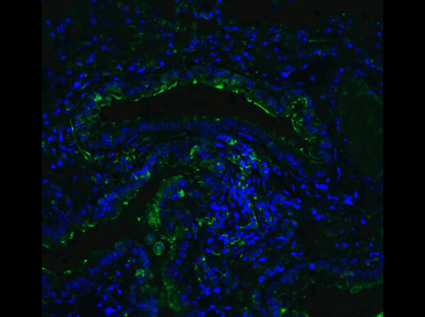
Immunofluorescence of Rabbit Anti-ACE2 Antibody. Tissue: Human Testis Tissue. Fixation: 4% PFA. Primary Antibody: Anti-ACE2 at 20µg/mL. Secondary Antibody: Goat anti-rabbit IgG secondary antibody at 1:500 dilution (green) and DAPI counterstain (blue).
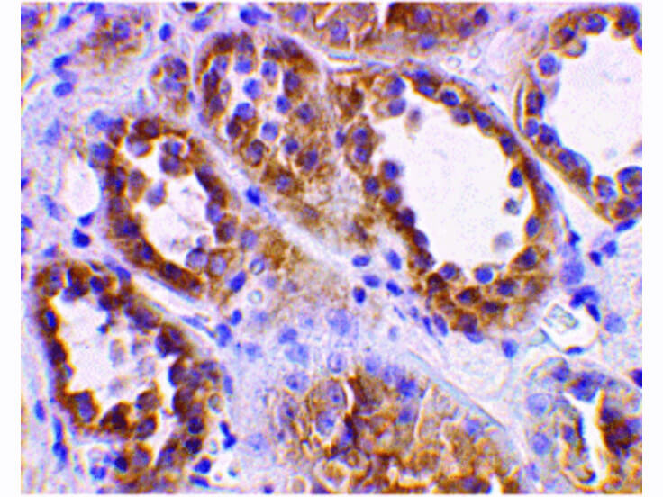
Immunocytochemistry of ACE2 antibody. Cell Type: human kidney cells. Fixation: formalin fixed paraffin embedded. Antigen retrieval: not required. Primary antibody: ACE2 antibody at 2 µg/mL for 1 h at RT. Secondary antibody: Peroxidase rabbit secondary antibody at 1:10,000 for 45 min at RT. Localization: ACE2 is located in the cell membrane and cytoplasm. Staining: ACE2 is stained with hematoxylin purple nuclear counterstain.
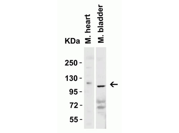
Western Blot of Rabbit Anti-ACE2 Antibody. Lane 1: Mouse Heart Lysate. Lane 2: Mouse Bladder Lysate. Load: 15µg of lysates per lane. Primary Antibody: Anti-ACE2 at 2µg/mL for 1hr at RT in 5% BLOTTO/TBST. Secondary Antibody: Goat anti-rabbit IgG HRP conjugate at 1:10000 dilution. Predicted MW: ~93kDa.
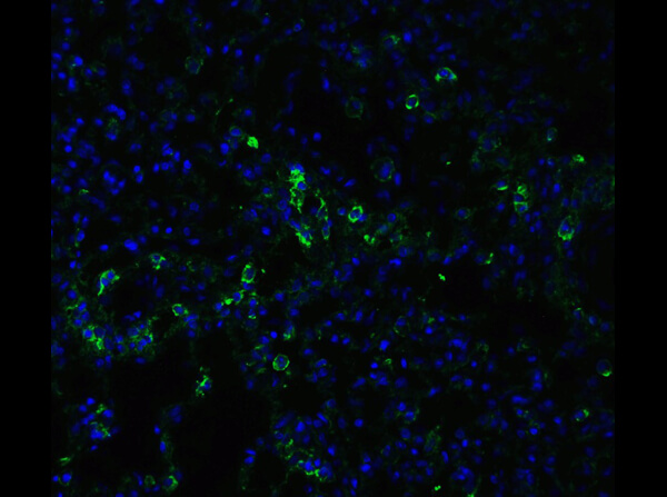
Immunofluorescence of Rabbit Anti-ACE2 Antibody. Tissue: Mouse Lung Tissue. Fixation: 4% PFA. Primary Antibody: Anti-ACE2 at 20µg/mL. Secondary Antibody: Goat anti-rabbit IgG secondary antibody at 1:500 dilution (green) and DAPI counterstain (blue).
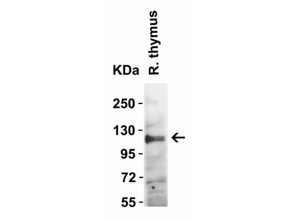
Western Blot of Rabbit Anti-ACE2 Antibody. Lane 1: Rat Thymus Lysate. Load: 15µg of lysate. Primary Antibody: Anti-ACE2 at 2µg/mL for 1hr at RT in 5% BLOTTO/TBST. Secondary Antibody: Goat anti-rabbit IgG HRP conjugate at 1:10000 dilution. Predicted MW: ~93kDa.
Immunofluorescence of Rabbit Anti-ACE2 Antibody. Tissue: Rat Lung Tissue. Fixation: 4% PFA. Primary Antibody: Anti-ACE2 at 20µg/mL. Secondary Antibody: Goat anti-rabbit IgG secondary antibody at 1:500 dilution (green) and DAPI counterstain (blue).
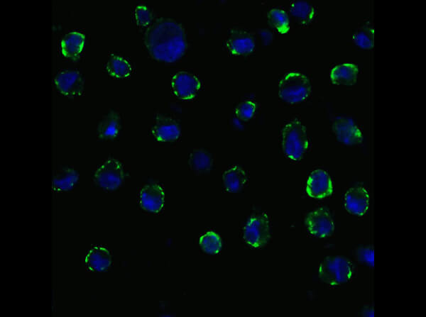
Immunofluorescence of Rabbit Anti-ACE2 Antibody. Tissue: Caco2 Cells. Fixation: 4% PFA. Primary Antibody: Anti-ACE2 at 20µg/mL. Secondary Antibody: Goat anti-rabbit IgG secondary antibody at 1:500 dilution (green) and DAPI counterstain (blue). Image showing membrane staining on Caco2 cells.
* Mehrwertsteuer und Versandkosten nicht enthalten. Irrtümer und Preisänderungen vorbehalten