Anti-ACE2 antibody was prepared from whole rabbit serum produced by repeated immunizations with a synthetic peptide corresponding to amino acids near the internal region of human ACE2.
Konjugation:
Unconjugated
Alternative Synonym:
ACE2 Antibody, ACEH, Angiotensin-converting enzyme 2, ACE-related carboxypeptidase, ACEH
Anti-ACE2 Antibody has been tested for use in ELISA, Immunofluorescence, Immunohistochemistry and Western Blotting. IHC and WB validated in human, mouse, and rat samples, IF validated in human samples. Expect a band at ~93 kDa in size corresponding to AC
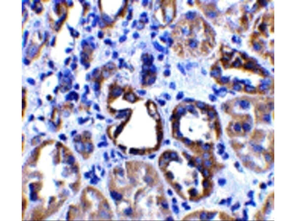
Immunohistochemistry of ACE2 Antibody. Tissue: paraffin-embedded human kidney tissue. Fixation: formaldehyde and blocked with 10% serum for 1 h at RT. Antigen retrieval: heat mediation with a citrate buffer (pH6). Primary Antibody: Anti-ACE2 at 2µg/ml overnight at 2-8C. Secondary Antibody: Goat anti-rabbit IgG H&L (HRP) at 1:250 secondary. Counter stained with Hematoxylin.
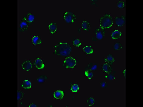
Immunofluorescence of Anti-ACE2 Antibody. Tissue: Caco2 Cells. Fixation: 4% PFA. Primary Antibody: Anti-ACE2 at 20µg/mL. Secondary Antibody: Goat anti-rabbit IgG secondary antibody at 1:500 dilution (green) and DAPI counterstain (blue). Image showing membrane staining on Caco2 cells.
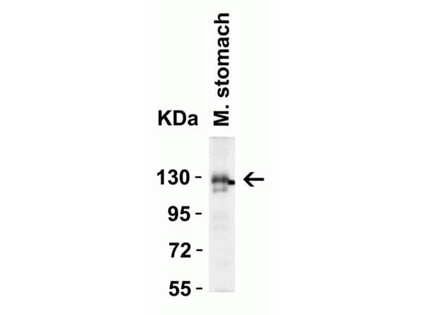
Western Blot of Rabbit anti-ACE2 antibody. Lane 1: Mouse Stomach Lysate. Load: 15 µg per lane. Primary antibody: ACE2 antibody 4µg/mL for 1hr at RT. Secondary antibody: Goat anti-Rabbit secondary HRP antibody at 1:10000 dilution. Block: 5% BLOTTO/TBST. Predicted MW: ~93kDa. Observed MW: ~130kDa.
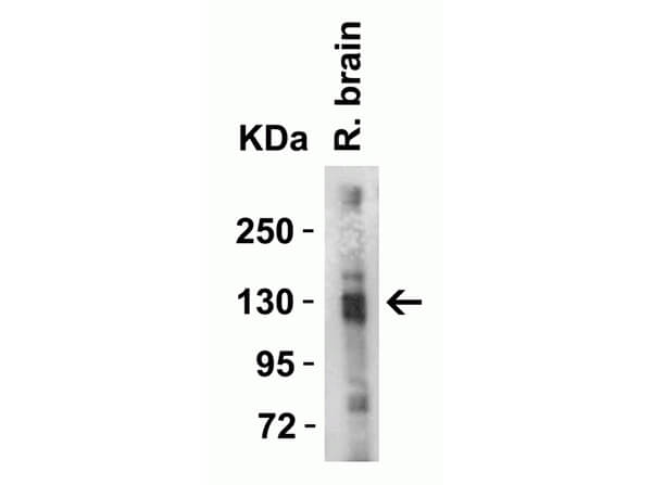
Western Blot of Rabbit anti-ACE2 antibody. Lane 1: Rat Brain Lysate. Load: 15 µg per lane. Primary antibody: ACE2 antibody 4µg/mL for 1hr at RT. Secondary antibody: Goat anti-Rabbit secondary HRP antibody at 1:10000 dilution. Block: 5% BLOTTO/TBST. Predicted MW: ~93kDa. Observed MW: ~130kDa.
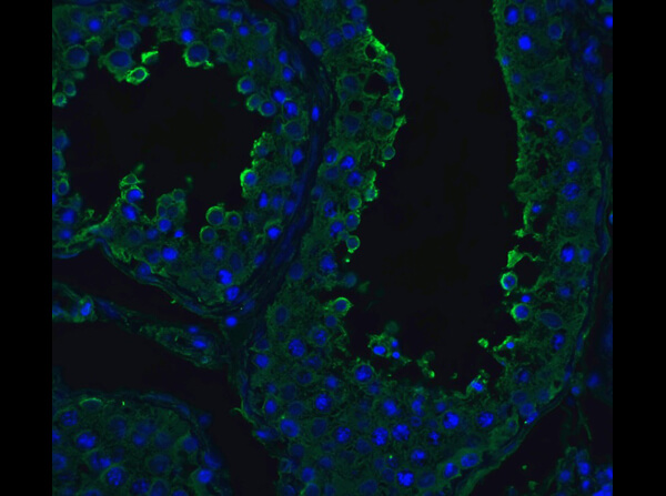
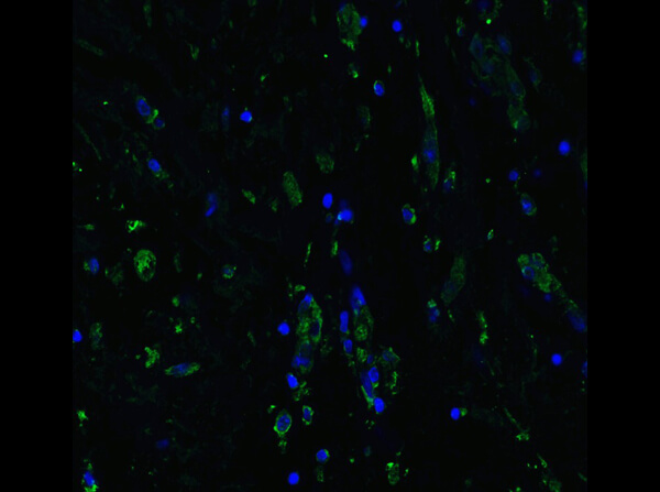
Immunofluorescence of Anti-ACE2 Antibody. Tissue: Human Testis Tissue. Fixation: 4% PFA. Primary Antibody: Anti-ACE2 at 20µg/mL. Secondary Antibody: Goat anti-rabbit IgG secondary antibody at 1:500 dilution (green) and DAPI counterstain (blue).
Western Blot of ACE2 antibody. Load: human kidney lysate 35 µg per lane. Primary antibody: ACE2 antibody at 2 µg/ml for overnight at 4C. Secondary antibody: Peroxidase rabbit secondary antibody at 1:10,000 for 45 min at RT. Block: 5% BLOTTO overnight at 4C. Predicted/Observed size: 92 kDa, 95 kDa for ACE2. Other band(s): ACE2 splice variants and isoforms.
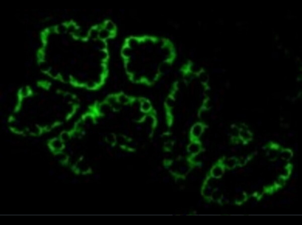
Immunofluorescence of Anti-ACE2 Antibody. Tissue: Human Kidney Tissue. Fixation: 4% PFA. Primary Antibody: Anti-ACE2 at 20µg/mL. Secondary Antibody: Goat anti-rabbit IgG secondary antibody at 1:500 dilution (green).
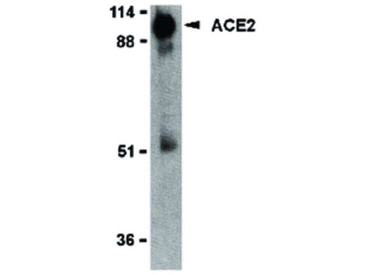
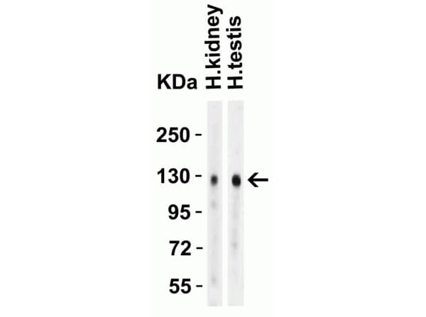
Western Blot of Rabbit anti-ACE2 antibody. Lane 1: Human Kidney Lysate. Lane 2: Human Testis Lysate. Load: 15 µg per lane. Primary antibody: ACE2 antibody 4µg/mL for 1hr at RT. Secondary antibody: Goat anti-Rabbit secondary HRP antibody at 1:10000 dilution. Block: 5% BLOTTO/TBST. Predicted MW: ~93kDa. Observed MW: ~130kDa.
Immunofluorescence of Anti-ACE2 Antibody. Tissue: Human Lung Tissue. Fixation: 4% PFA. Primary Antibody: Anti-ACE2 at 20µg/mL. Secondary Antibody: Goat anti-rabbit IgG secondary antibody at 1:500 dilution (green) and DAPI counterstain (blue).
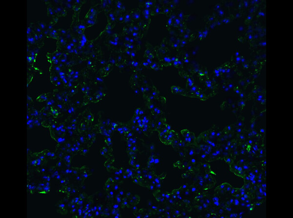
Immunofluorescence of Anti-ACE2 Antibody. Tissue: Mouse Lung Tissue. Fixation: 4% PFA. Primary Antibody: Anti-ACE2 at 20µg/mL. Secondary Antibody: Goat anti-rabbit IgG secondary antibody at 1:500 dilution (green) and DAPI counterstain (blue).
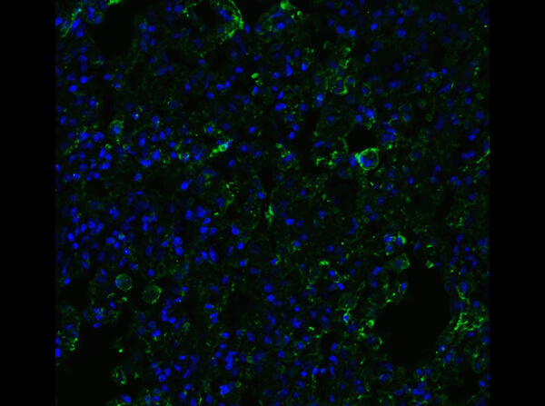
Immunofluorescence of Anti-ACE2 Antibody. Tissue: Rat Lung Tissue. Fixation: 4% PFA. Primary Antibody: Anti-ACE2 at 20µg/mL. Secondary Antibody: Goat anti-rabbit IgG secondary antibody at 1:500 dilution (green) and DAPI counterstain (blue).
* Mehrwertsteuer und Versandkosten nicht enthalten. Irrtümer und Preisänderungen vorbehalten