Anti-Ambra1 antibody was prepared from whole rabbit serum produced by repeated immunizations with a 15 amino acid synthetic peptide from near the C-terminus of human Ambra1.
Konjugation:
Unconjugated
Alternative Synonym:
Ambra1 Antibody, DCAF3, WDR94, KIAA1736, Activating molecule in BECN1-regulated autophagy protein 1
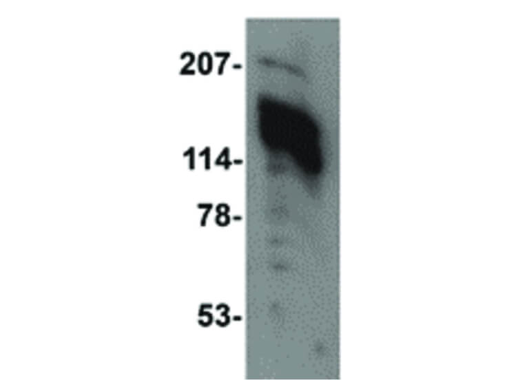
Anti-Ambra1 Antibody has been tested for use in ELISA, Western Blotting, Immunohistochemistry and Immunofluorescence. Specific conditions for reactivity should be optimized by the end user. Expect a band at approximately 143 kDa in Western Blots of speci
Western Blot of Ambra1 antibody. Lane 1: 3T3 cell lysate at 1 µg/mL. Load: 35 µg per lane. Secondary antibody: Peroxidase rabbit secondary antibody at 1:10,000 for 45 min at RT. Block: 5% BLOTTO overnight at 4C. Predicted/Observed size: 142.5 kDa, ~140 kDa for Ambra1.
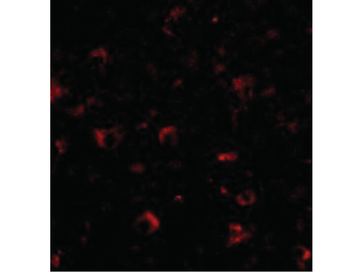
Immunofluorescence Microscopy of Ambra1 antibody. Tissue: Human brain cells. Fixation: 0.5% PFA. Antigen retrieval: not required. Primary antibody: Ambra1 antibody at 20 µg/mL for 1 h at RT. Secondary antibody: Fluorescein rabbit secondary antibody at 1:10,000 for 45 min at RT. Staining: Ambra1 as a red fluorescent signal.
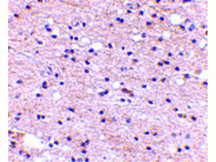
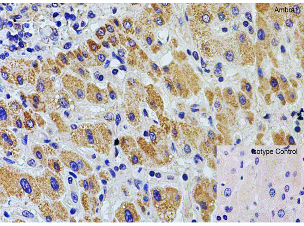
Immunohistochemistry of Ambra1 antibody. Tissue: Human brain tissue. Fixation: formalin fixed paraffin embedded. Antigen retrieval: not required. Primary antibody: Ambra1 antibody at 5 µg/mL for 1 h at RT. Secondary antibody: Peroxidase rabbit secondary antibody at 1:10,000 for 45 min at RT. Localization: Ambra1 is nuclear and occasionally cytoplasmic. Staining: Ambra1 as a precipitated red signal with hematoxylin purple nuclear counterstain.
Immunofluorescence of Ambra1. Cell: K562 cells. Primary Antibody: Ambra1 Antibody at 20 µg/ml.
Immunohistochemical of Ambra1. Tissue: rat colon tissue. Fixation: paraffin-embedded, formaldehyde, and blocked with 10% serum for 1 h at RT. Antigen retrieval: heat mediation with a citrate buffer (pH6). Primary Antibody: anti-AMBRA1 antibody at 2 ug/ml overnight at 4C. Secondary: goat anti-rabbit IgG H&L (HRP) at 1:250. Counter stained with Hematoxylin.
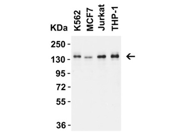
Western Blot Validation of Ambra1. Load: 15µg of human cell lysate per lane. Lane 1: K562, Lane 2: MCF7, Lane 3: Jurkat, Lane 4: THP-1. Primary Antibody: AMBRA1 at 1 ug/mL for 1 h incubation at RT in 5% NFDM/TBST. Secondary: Goat Anti-Rabbit IgG HRP conjugate at 1:10000 dilution.
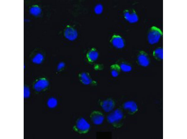
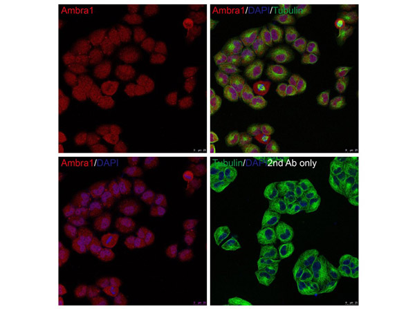
Immunofluorescent of Ambra1. Fixation: PFA-fixed HeLa cells. Primary Antibody: AMBRA1 at 20 ug/mL. Secondary: goat anti-rabbit IgG antibody at 1:1000 dilution (red) and DAPI staining (blue). Alpha tubulin was stained with anti-alpha tubulin antibody following by goat anti-mouse IgG secondary antibody (green). Images were captured with confocal microscopy.
* Mehrwertsteuer und Versandkosten nicht enthalten. Irrtümer und Preisänderungen vorbehalten