Anti-BCMA antibody was prepared from whole rabbit serum produced by repeated immunizations with a synthetic peptide mapping at the C-terminus of human BCMA. The immunogen is located within the last 50 amino acids of BCMA.
Konjugation:
Unconjugated
Alternative Synonym:
BCMA Antibody, BCM, BCMA, CD269, TNFRSF13A, BCM, Tumor necrosis factor receptor superfamily member 17, B-cell maturation protein
Anti-BCMA Antibody has been tested for use in ELISA, Western Blotting, Immunohistochemistry and Immunofluorescence. Specific conditions for reactivity should be optimized by the end user. Expect a band at approximately 20 kDa in Western Blots of specific
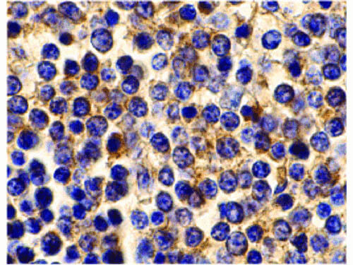
Immunohistochemistry of BCMA antibody. Tissue: Human spleen cells. Fixation: formalin fixed paraffin embedded. Antigen retrieval: not required. Primary antibody: BCMA antibody at 10 µg/mL for 1 h at RT. Secondary antibody: Peroxidase rabbit secondary antibody at 1:10,000 for 45 min at RT. Localization: BCMA is localized in the cell membrane and in the endomembrane system. Staining: BCMA as precipitated brown signal with hematoxylin blue nuclear counterstain.
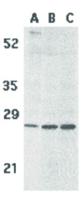
Western Blot of BCMA antibody. Lane 1: Human spleen tissue lysate. Lane 2: K562 cell lysate. Lane 3: U937 cell lysate. Load: 35 µg per lane. Primary antibody: BCMA antibody at 5 µg/mL for overnight at 4C. Secondary antibody: Peroxidase rabbit secondary antibody at 1:10,000 for 45 min at RT. Block: 5% BLOTTO overnight at 4C. Predicted/Observed size: 20.2 kDa, 28 kDa for BCMA. Other band(s): BCMA splice variants and isoforms.
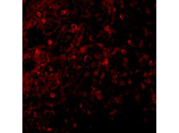
Immunofluorescence Validation of BCMA. Tissue: Human Spleen. Fixation: 4% paraformaldehyde-fixed. Primary: BCMA at 10 µg/mL, followed by goat anti-rabbit IgG secondary antibody at 1:500 dilution (red).
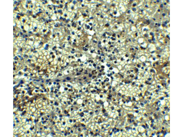
Immunohistochemistry Validation of BCMA. Tissue: Human Spleen. Fixation: paraffin-embedded, formaldehyde and blocked with 10% serum for 1 h at RT. Antigen retrieval: heat mediation with a citrate buffer (pH6). Primary Antibody: anti-BCMA antibody at 5 µg/ml overnight at 4C. Secondary: goat anti-rabbit IgG H&L (HRP) at 1:250. Counter stained with Hematoxylin.
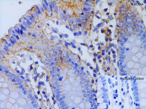
Immunohistochemistry Validation of BCMA.Tissue: Human Colon. Fixation: paraffin-embedded, formaldehyde and blocked with 10% serum for 1 h at RT. Antigen retrieval: heat mediation with a citrate buffer (pH6). Primary Antibody: anti-BCMA antibody at 1 µg/ml overnight at 4C. Secondary: goat anti-rabbit IgG H&L (HRP) at 1:250. Counter stained with Hematoxylin.
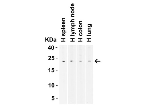
Western Blot Validation of BCMA.Load: 10 µg of human lysates. Lane 1: human spleen, Lane 2: human lymph node, Lane 3: human colon, Lane 4: human lung. Primary Antibody: BCMA 2 µg/mL for 1 h incubation at RT in 5% NFDM/TBST. Secondary: Goat Anti-Rabbit IgG HRP conjugate at 1:10000 dilution.
* Mehrwertsteuer und Versandkosten nicht enthalten. Irrtümer und Preisänderungen vorbehalten