Collagen Type I Antibody Biotin Conjugated, Rabbit, Polyclonal
Artikelnummer:
ROC-600-406-103
Hersteller Artikelnummer:
600-406-103
Alternativnummer:
ROC-600-406-103
Hersteller:
Rockland Immunochemicals
Wirt:
Rabbit
Kategorie:
Antikörper
Applikation:
DOT, FC, Multiplex Assay
Spezies Reaktivität:
Human, Mouse, Rat
Immunogen:
Collagen Type I from human and bovine placenta.
Konjugation:
Biotin
Alternative Synonym:
rabbit anti-collagen type I antibody biotin conjugation, biotin conjugated rabbit anti-collagen type I antibody, Collagen Of Skin Tendon And Bone, Collagen Type 1 antibody, Collagen type I alpha 1 antibody, Collagen alpha-1 (I) chain, Alpha-1 type I collagen, type 1 procollagen alpha 1
Anti-COLLAGEN Type I Antibody Biotin Conjugated has been tested by dot blot and Flow Cytometry and is suitable for western blot, immunoprecipitation, Flow Cytometry, and immunohistochemistry. Researchers should determine optimal titers for applications t
Flow Cytometry of Rabbit Anti-Collagen 1 Antibody. Cells: primary adult human dermal fibroblast cells. Stimulation: none. Primary antibody: Biotin-Conjugated Collagen 1 antibody (600-406-103) at 5µg/mL for 45 min at 4C. Secondary antibody: Rabbit Streptavidin, R-PE antibody at 1:500 for 15 min at RT. Courtesy of D. Figueroa NIH.
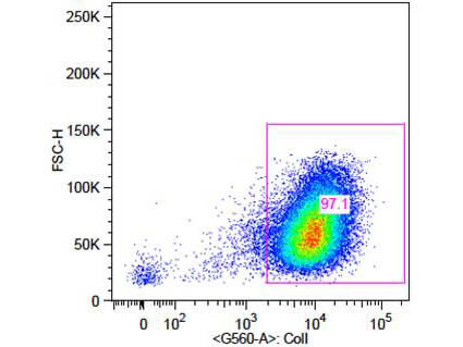
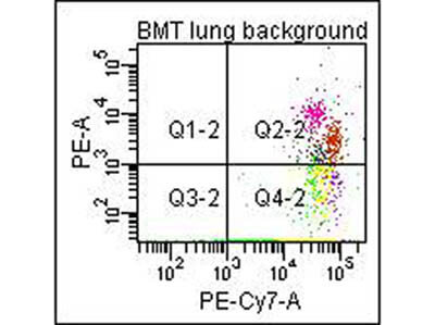
Flow Cytometry of Anti-Collagen Type I Biotin Conjugated Antibody (600-406-103). Showing a 7.4% of fibrocyte population in the mouse lung using CD45 and Col 1A1 double positive markers. In the experiment, we used Biotinated Collagen Type 1 (p/n 600-406-103) in combination with a PE-conjugated secondary antibody for flow cytometric analysis. Courtesy of Walden Ai, PhD, Univ of SC School of Medicine.
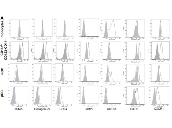
Flow cytometry of Anti-Collagen Type I Antibody Biotin Conjugated.Expanded CD11chiCD123-CD14-cells are fibrocytes that mediate angiogenesis.(A) Using the same gating strategy as shown inFigure 1A, CD11chiCD123-CD14-cells from a representative subject sample were analyzed for cell surface phenotype. The shaded areas represent background fluorescence on the designated population as indicated by FMO controls. This is representative of more than 10 experiments.Figure 2. PMID: 23757729.
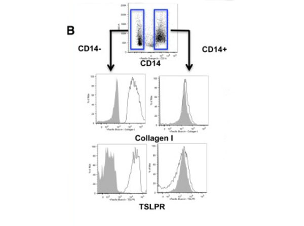
Flow Cytometry of Anti-Collagen Type I Antibody Biotin Conjugated.IL-4 induces monocytes to differentiate into CD14-fibrocytes that are readily distinguished from CD14+macrophages in the same culture. (B) Cell surface phenotype of IL-4-differentiated adherent cells identifies 2 subsets based on CD14 expression, which further shows differential expression of collagen and TSLPR. FMO controls on gated CD14+vs CD14-populations are shown by shaded gray histograms. This is representative of more than 5 experiments from 5 separate healthy donors.Figure 5. PMID: 23757729.
* Mehrwertsteuer und Versandkosten nicht enthalten. Irrtümer und Preisänderungen vorbehalten