Anti-Green Fluorescent Protein (GFP) is produced by immunizing GFP containing fusion protein that corresponds to the full length amino acid sequence (246aa) derived from the jellyfish Aequorea victoria.
Konjugation:
Biotin
Alternative Synonym:
rabbit anti-GFP antibody biotin conjugation, biotin conjugated rabbit anti-GFP antibody, Green Fluorescent Protein, GFP antibody, Green Fluorescent Protein antibody, EGFP, enhanced Green Fluorescent Protein, Aequorea victoria, Jellyfish
Biotin Conjugated GFP Antibody has been tested by ELISA and western blot and is suitable for immunoblotting, ELISA, immunohistochemistry, immunomicroscopy as well as other antibody based assays using streptavidin or avidin conjugates requiring lot-to-lot
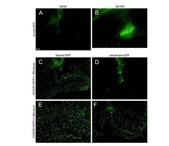
Immuno-Fluorescence of Biotin Mouse anti-GFP antibody used at 1:5000. Montage of microscopic fields 24 hours after Tat (Panels A-C, E) or control vehicle (Panels D, F) injection into the right hippocampus of irradiation chimeras. Images in Panels A, B, C, and D were taken from CD45.1 host mice engrafted with heterozygous CX3CR1/GFP+/- bone marrow, whereas images E and F were from CD45.1 host mice engrafted with homozygous CX3CR1/GFP+/+ bone marrow. A, C, D, E, and F were taken with a 4* objective with the same 0.9 sec exposure time and B was taken with a 10* objective with a longer exposure time (4 sec) to intensify the weak fluorescence of the GFP signal. Panel A and B depict very low expression of fluorescence from infiltrating GFP expressing leukocytes, while Panels C-F depict amplified GFP expression with anti-GFP antibody and Alexa 488 conjugated second antibody treatment. All experimental and control groups, n=3 independent replicates. Scale bar=100 µm for Panels A, C, D, E, and F, 40 µm for B. Fig 2. PMID: 21912650.
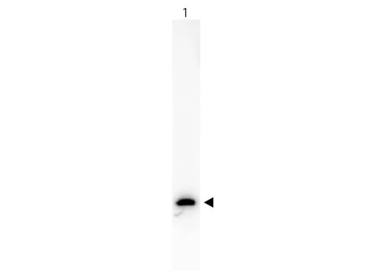
Western Blot of Peroxidase conjugated Goat anti-Rabbit IgG antibody. Lane 1: 50ng of GFP. Lane 2: none. Primary antibody: none. Secondary antibody: Anti-GFP Antibody Biotin Conjugated secondary antibody was used at 1:5000 in Blocking Buffer for Fluorescent Western Blotting (p/n MB-070) for 45 min at RT. HRP Streptavidin (p/n S000-03) was used at 1:40,000 in MB-070 for 30 min at 20C. Block: 5% Blotto (p/n B501-0500) 30 min at 20C. Predicted/Observed size: 28 kDa for GFP. Other band(s): none.
* Mehrwertsteuer und Versandkosten nicht enthalten. Irrtümer und Preisänderungen vorbehalten