MYC Epitope Tag Antibody Biotin Conjugated, Rabbit, Polyclonal
Artikelnummer:
ROC-600-406-381
Hersteller Artikelnummer:
600-406-381
Alternativnummer:
ROC-600-406-381
Hersteller:
Rockland Immunochemicals
Wirt:
Rabbit
Kategorie:
Antikörper
Applikation:
DOT, ELISA, WB
Immunogen:
This antibody was purified from whole rabbit serum prepared by repeated immunizations with Myc epitope tag peptide E-Q-K-L-I-S-E-E-D-L conjugated to KLH using maleimide. The sequence corresponds to aa 410-419 of human c-Myc.
Konjugation:
Biotin
Alternative Synonym:
Rabbit anti-MYC Epitope Tag Biotin Conjugated Antibody, Rabbit anti-c-myc tag Biotin conjugation, Glu-Gln-Lys-Leu-Ile-Ser-Glu-Glu-Asp-Leu
Klonalität:
Polyclonal
Konzentration:
1.0 mg/mL by UV absorbance at 280 nm
Puffer:
0.02 M Potassium Phosphate, 0.15 M Sodium Chloride, pH 7.2
Anti-myc has utility to detect the fusion protein of the myc epitope cloned along with the target gene. As such, anti-myc/myc can be used to identify fusion proteins containing the myc epitope. The antibody recognizes the Myc tag fused either to the amin
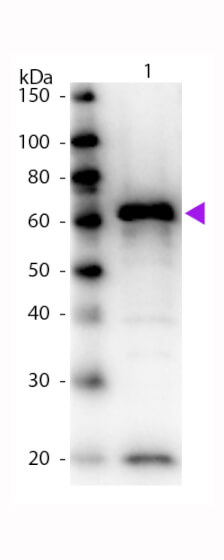
Western Blot of Biotin Conjugated Rabbit anti-Myc Epitope Tag antibody. Lane 1: 12-Tag Lysate. Lane 2: None. Load: N/A. Primary antibody: None. Secondary antibody: Biotin rabbit secondary antibody at 1:1,000 for 60 min at RT. Block: MB-070 for 30 min at RT. Predicted/Observed size: 60 kDa, 60 kDa for Myc Epitope Tag. Other band(s): Myc splice variants and isoforms.
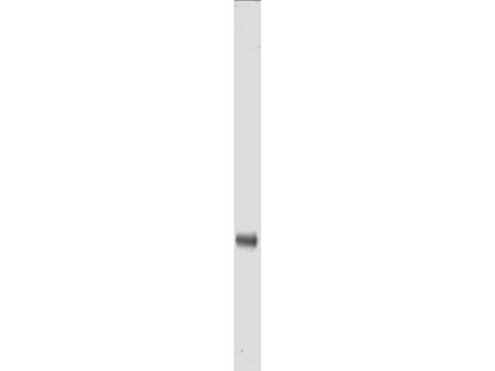
Anti-Myc epitope tag polyclonal antibody detects Myc-tagged recombinant proteins by western blot. Polyclonal rabbit-anti-Myc epitope tag at 0.5-1.0 µg/ml was used to detect 1.0 ug of recombinant protein containing the Myc epitope tag. A 4-20% gradient gel was used to separate the protein by SDS-PAGE. The protein was transferred to nitrocellulose using standard methods. After blocking the membrane was probed with the primary antibody for 1 h at room temperature followed by washes and reaction with a 1:2500 dilution of IRDye(TM)800 conjugated Gt-a-Rabbit IgG [H&L] (code 611-132-122) for 30 min at room temperature. LICORs Odyssey Infrared Imaging System was used to scan and process the image. Other detection systems will yield similar results.
* Mehrwertsteuer und Versandkosten nicht enthalten. Irrtümer und Preisänderungen vorbehalten