The immunogen is a Red Fluorescent Protein (RFP) fusion protein corresponding to the full length amino acid sequence (234aa) derived from the mushroom polyp coral Discosoma.
Konjugation:
Unconjugated
Alternative Synonym:
chicken anti-RFP antibody, DsRed, rDsRed, Discosoma sp. Red Fluorescent Protein, Red fluorescent protein drFP583
0.02 M Potassium Phosphate, 0.15 M Sodium Chloride, pH 7.2
Formulierung:
Liquid (sterile filtered)
Antibody Type:
Primary Antibody
Application Verdünnung:
ELISA: 1:10,000, WB: 1:1,000 - 1:3,000
Anwendungsbeschreibung:
Anti-RFP is designed to detect recombinant RFP. Anti-RFP antibody has been tested by ELISA and western blot to detect RFP. Use either alkaline phosphatase or peroxidase conjugated polyclonal anti-RFP to detect RFP or RFP containing proteins on western bl
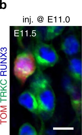
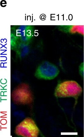
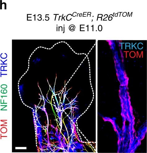
PSNs with high TRKC levels preferentially survive the cell death period. a Temporal fate mapping of TRKCHigh PSNs by 4-OHT (low dose, 0.02g/kg). b-d Injection of TrkCCreER,R26tdTOM mice with low dose of 4-OHT at E11.0, DRG analyzed at E11.5 with recombination in few (b), preferentially high TRKC PSNs (c, d) (P<0.001). Frequency distribution of TOM+ PSNs according to TRKC intensity (c) and pie charts (d) illustrating the large proportion of TOM+ cells among TRKCHigh PSNs. Scale bar: 50µm. e, f Percentage of recombined PSNs at E11.5 and E13.5 in DRGs from TrkCCreER,R26tdTOM animals after 4-OHT
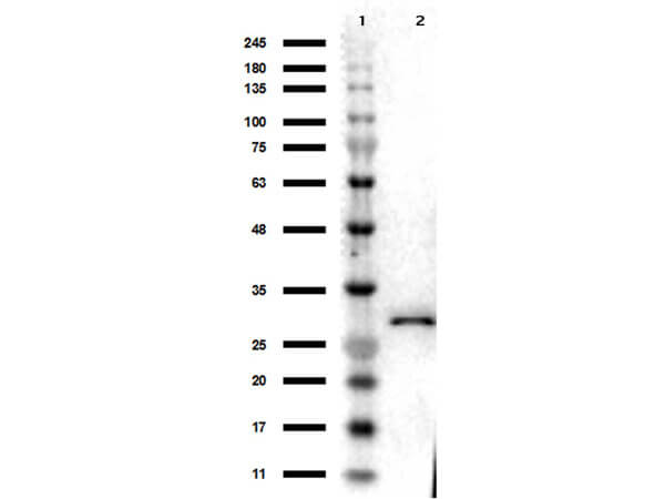
Western Blot Results of Chicken Anti-RFP Antibody. Lane 1: Opal PreStained Molecular Weight Marker (p/n MB-210-0500). Lane 2: RFP (p/n 000-001-379), load 50ng. Primary Antibody: Anti-RFP 1µg/mL overnight at 4C. Secondary Antibody: Goat Anti-Chicken HRP (p/n 603-103-126) at 1:40,000 for 30min at RT. Blocking: BlockOut (p/n MB-073) for 30min at RT. Expect: 27kDa.
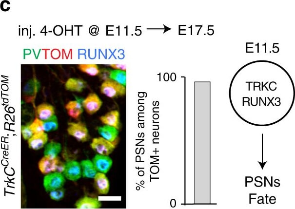
Differential expression of TRKC in PSNs prior to the cell death period. a Scheme of our working hypothesis. b, c Temporal fate mapping of TRKC PSNs by 4-OHT induction. TrkCCreER mice allow temporary activation of CreER in the TRKC+ cells 2h after 4-OHT injection21,22. Immunostaining for PV, RFP and RUNX3 on E17.5 DRG sections (c) and graph showing distribution of PV+/RUNX3+ PSNs among the TOM+ cells (n=4). Scale bar: 20µm. d Quantification of PSNs at C5 and C7. ***P<0.001, one-way analysis of variance (ANOVA) with Sidaks multiple comparisons test (n=2-3). The window of PSNs cell death is shown. e TRKC expression in E11.5 ISL1+ (and RUNX3+, whose staining is not shown for more visibility) DRG neurons. Scale bar: 50µm. f TRKC levels in PSNs of eillustrated by color coding, dark blue indicates the lower and red the higher TRKC levels. From here, all observations are done at brachial levels (C5-8). g Distribution of TRKC levels in PSNs from e. h Distribution of TRKC levels in PSNs in E11.5 DRG neurons (from g). The data exhibit a Poisson-like distribution (one representative animal), with the mean used to define the two different categories of TRKC intensity (TRKCHigh and TRKCLow). i Projection of seven images of RUNX3+/TRKC+ PSNs from one brachial DRG, dots indicate TRKC-labeled neurons and color codes reveal TRKC intensity as shown in h. j Projection image of smFISH for pan Ntrk3 and Ntrk3 full length (FL) transcripts in E11.5 DRG, visualized at high magnification in (1) and (2) (images show full projection), right panel shows color coding of Ntrk3 FL levels in red, the brighter, the higher levels. k Distribution of the number of Ntrk3 FL molecules in E11.5 DRG neurons by smFISH, normalized to pan Ntrk3 (Ntrk3 FL represent 68% of all Ntrk3 transcripts). lTrkCCreER,R26tdTOM mice were injected at E9.75 with 4-OHT and analyzed at E11.5 (n=3). m, n Frequency distribution (m) and pie chart (n) of TOM+/TRKC+ neurons from l according to their level of TRKC intensity. Source data are available as a Source Data file Figure provided by CiteAb. Source: Nat Commun, PMID: 31515492.
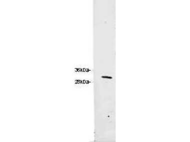
Western blot of Rocklands Chicken Anti-RFP Antibody. Loaded 0.1 µg of RFP protein (p/n 000-001-379) on a 4-20% gel and transferred to nitrocellulose membrane. Chicken anti-RFP Antibody was added at 1.0 µg/mL at RT for 2 hours. IRDye800 goat anti-Chicken (p/n 603-132-126) was added at 1:20,000 at RT for 45 minutes.
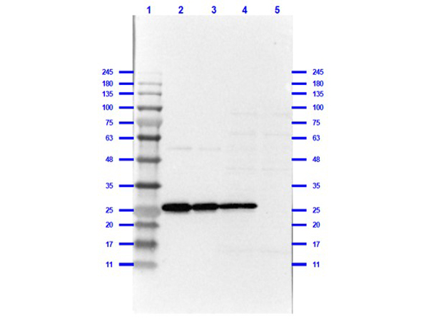
Western Blot of Chicken Anti-RFP Antibody. Lane 1: Prestained MW marker (p/n MB-210-0500). Lane 2: RFP (p/n 000-001-379) 0.1µg reduced [+]. Lane 3: RFP (p/n 000-001-379) 0.05µg reduced [+]. Lane 4: RFP/HEK293T (p/n 000-001-379/W09-001-GX5) 0.5µg/10µg reduced [+]. Lane 5: HEK293T (p/n W09-001-GX5) 10µg reduced [-]. Blocking Buffer: BlockOut Universal Blocking Buffer (p/n MB-073) for 1hr at RT. Primary Antibody: Chicken Anti-RFP at 1µg/mL overnight at 2-8C. Secondary Antibody: Rabbit anti-Chicken IgG HRP (p/n 603-4302) at 1:40,000 for 30 mins at RT. Predicted/Observed: 27kda.
* Mehrwertsteuer und Versandkosten nicht enthalten. Irrtümer und Preisänderungen vorbehalten