0.02 M Potassium Phosphate, 0.15 M Sodium Chloride, pH 7.2
Formulierung:
Lyophilized
Target-Kategorie:
Guinea Pig
Antibody Type:
Secondary Antibody
Application Verdünnung:
FLISA: >1:20,000, IF Microscopy: >1:5,000, WB: >1:10,000
Anwendungsbeschreibung:
The emission spectra for this DyLight(TM) conjugate match the principle output wavelengths of most common fluorescence instrumentation. This product is designed for immunofluorescence microscopy, fluorescence based plate assays (FLISA) and fluorescent weste
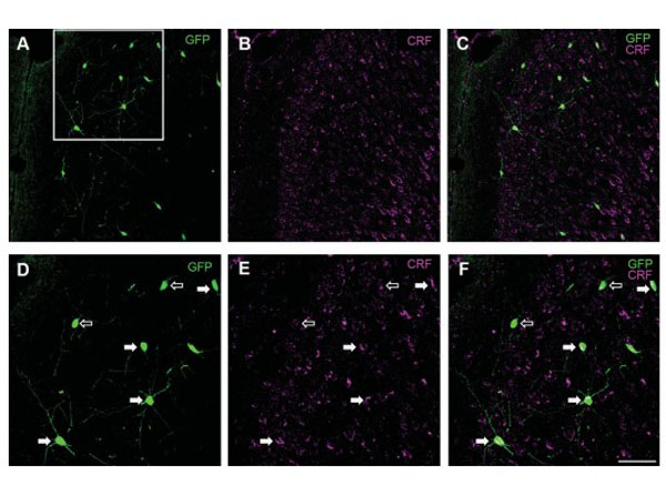
Analysis of corticotropin-releasing factor (CRF)-expressing interneurons in the cingulate cortex of FVB-Tg(GadGFP)45704Swn/J mice. A-C: Representative confocal images of cells in the cingulate cortex immunopositive for GFP (green) and CRF (magenta). The white square in A represents the area that is shown at higher magnification in D-F. D-F: Detailed analysis of CRF expression in GFP 1 cells in the cingulate gyrus. Solid white arrows indicate GFP1/CRF 1 cells and open white arrows GFP1/CRF2 cells. Note the widespread CRF 1 punctae throughout cells in layers II-III of the cingulate cortex. Scale bar in F 5 50 lm for A-C, 25 lm for D-F. Figure 9. PMID: .26669716
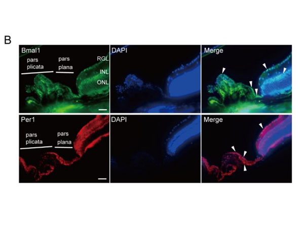
Immunohistochemical analysis in the retina and ciliary body of C57BL/6J mice. (A) Adrenergic beta2-receptors (Adrb2) and glucocorticoid receptor (GR) immunoreactivity in the retina and ciliary body of C57BL/6J mice. (B) Immunoreactivity of clock protein Bmal1 and Per1 in the retina and ciliary body of C57BL/6J mice. Fluorescence immunohistochemistry revealed that immunoreactivity (white arrowhead) localized in the epithelia of the ciliary body. Scale bar: 100 µm. DAPI, 4,6-diamidino-2-phenylindole. RGL, retinal ganglion cell layer, INL, inner nuclear layer, ONL, outer nuclear layer. Fig. S4. PMID: 32182332.
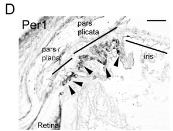
Localizations of GR, Adrb2, and Bmal1 in the ciliary body. Representative images of (A) GR, (B) adrenergic beta2-receptors (Adrb2), (C) Bmal1, and (D) Per1 immunoreactivity in the retina and ciliary body of albino Balb/c mice by immunohistochemistry. Strong immunoreactivity (arrowhead) was seen in the pars plana of the ciliary body. Scale bar: 100µm. CPE, ciliary pigmented epithelium, INL, inner nuclear layer, ONL, outer nuclear layer, RGL, retinal ganglion cell layer, TM, trabecular meshwork. Figure 3. PMID: 32182332.
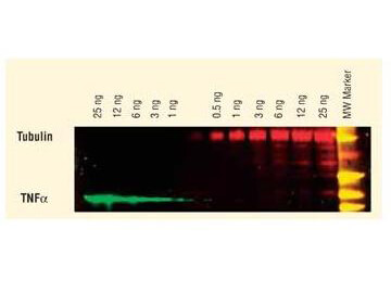
DyLight(TM) dyes can be used for two-color western blot detection with low background and high signal. Anti-tubulin was detected using a DyLight(TM) 549 conjugate. Anti-TNFa was detected using a DyLight(TM) 649 conjugate. The image was captured using the Typhoon(TM) 9410 Imaging System.
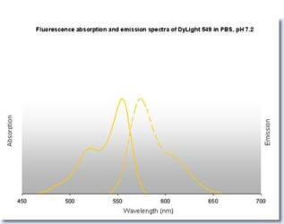
DyLight(TM) 549 Fluorescence Spectra.
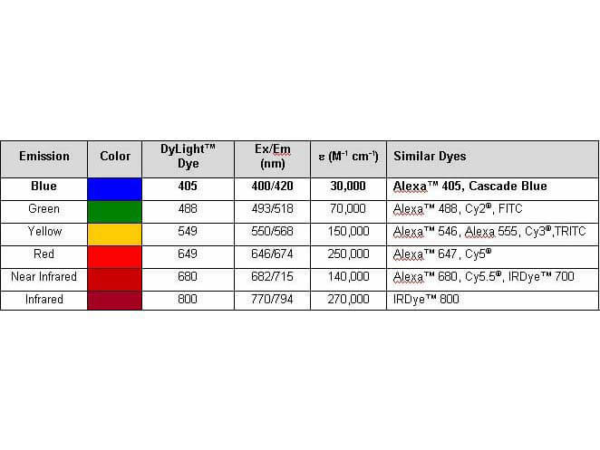
Properties of DyLight(TM) Conjugates.
* Mehrwertsteuer und Versandkosten nicht enthalten. Irrtümer und Preisänderungen vorbehalten