0.02 M Potassium Phosphate, 0.15 M Sodium Chloride, pH 7.2
Formulierung:
Lyophilized
Target-Kategorie:
Mouse
Antibody Type:
Secondary Antibody
Application Verdünnung:
FLISA: >1:20,000, IF Microscopy: >1:5,000, WB: >1:10,000
Anwendungsbeschreibung:
This product is designed for immunofluorescence microscopy, fluorescence based plate assays (FLISA) and fluorescent western blotting. This product is also suitable for multiplex analysis, including multicolor imaging, utilizing various commercial platfor
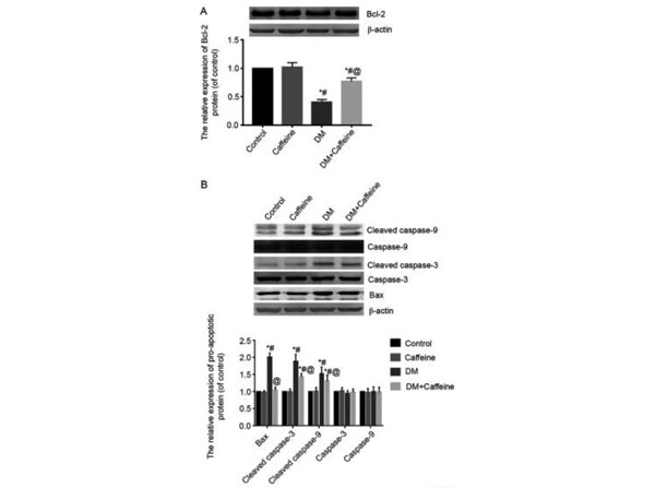
Western Blot Results using Goat Anti-Mouse IgG (H&L) Antibody DyLight(TM) 680 Conjugated. Expression levels of Bcl-2, Bax, caspase-3 and caspase-9 proteins in the DRG. (A) The expression level of Bcl-2 protein in rat bladders was detected by western blot analysis. (B) The expression levels of Bax, caspase-3, cleaved caspase-3, caspase-9 and cleaved caspase-9 proteins in rat bladders were detected by western blot analysis.*P<0.05 vs. control group,P<0.05 vs. caffeine group, P<0.05 vs. DM group. Bcl-2, B-cell lymphoma-2, Bax, Bcl-2-associated X protein, DRG, dorsal root ganglion, DM, diabetes mellitus. Fig 3. PMID: 33791010.
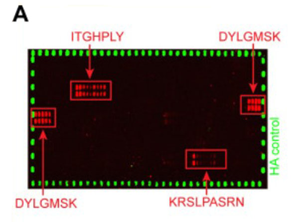
Peptide array results using Goat Anti-Mouse IgG (H&L) Antibody DyLight(TM) 680 Conjugated. Peptide arrays identify known immunogenic epitopes in L1.(A) Synthetic 15-mer peptides with residue overlaps of 14 residues were spotted on microarrays and incubated with serum mix from five tumor-bearing animals with high titers against both L1 isoforms. Bound serum antibodies were detected with fluorophore-conjugated secondary antibodies. Fig 5. PMID: 32746966.
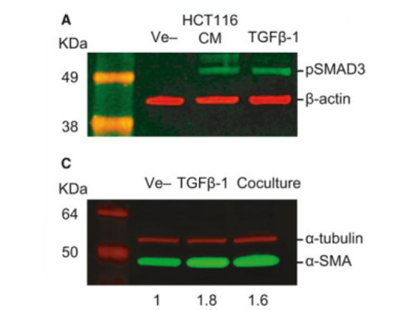
Western Blot Results using Goat Anti-Mouse IgG (H&L) Antibody DyLight(TM) 680 Conjugated. TGF-beta-mediated crosstalk between pericytes and CRC cells modulates pericyte secretome. (A) Incubation in HCT116 CM for 1h induces SMAD3 phosphorylation in PC, as assessed by western blot. Exogenous recombinant TGF-beta (10ngmL-1) was used as a positive control, and beta-actin was used as loading control (n=3). (B) Confocal microscopy images of SMAD3 subcellular localization in PC cultured alone or cocultured with HCT116 cells for 48h (n=3). SMAD3 is detected in the cytoplasm of PC in monoculture (arrows show nonstained nuclei). Nuclear translocation of SMAD3 takes place after coculture with HCT116 cells for 48h (arrowheads indicate stained nuclei). HCT116 cells treated with 10ngmL-1TGF-beta1 were used as a positive control. Scale bar=10µm. Fig 5. PMID: 32767843.
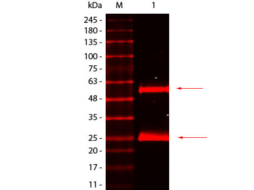
Western Blot of Goat anti-Mouse IgG Antibody DyLight 680 Conjugated Pre-absorbed. Lane 1: Mouse IgG. Load: 50 ng per lane. Primary antibody: none. Secondary antibody: Goat anti-Mouse IgG Antibody DyLight 680 Conjugated Pre-absorbed at 1:1,000 for 60 min at RT. Block: MB-070 for 30 min at RT. Predicted/Observed size: 55 kDa, 25 kDa for Mouse IgG.
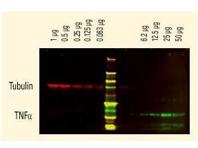
DyLight(TM) dyes can be used for two-color Western Blot detection with low background and high signal. Anti-tubulin was detected using a DyLight(TM) 680 conjugate. Anti-TNFa was detected using a DyLight(TM) 800 conjugate. The image was captured using the Odyssey Infrared Imaging System developed by LI-COR.
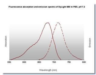
DyLight(TM) 680 Fluorescence Spectra
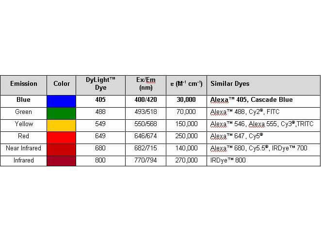
Properties of DyLight(TM) Conjugates.
* Mehrwertsteuer und Versandkosten nicht enthalten. Irrtümer und Preisänderungen vorbehalten