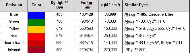
Mouse IgG Fc Antibody DyLight(TM) 405 Conjugated, DL405, Goat, Polyclonal
Artikelnummer:
ROC-610-146-003
Hersteller Artikelnummer:
610-146-003
Alternativnummer:
ROC-610-146-003
Hersteller:
Rockland Immunochemicals
Wirt:
Goat
Kategorie:
Antikörper
Spezies Reaktivität:
Mouse
Immunogen:
Mouse IgG F(c) fragment
Konjugation:
DL405
Alternative Synonym:
Goat Anti Mouse IgG F(c) Antibody DyLight(TM) 405 Conjugated, Goat Anti-Mouse IgG Fc Antibody DyLight(TM) 405 Conjugated, Goat Anti Mouse IgG Fc Fragment Antibody DyLight(TM) 405 Conjugated
Klonalität:
Polyclonal
Konzentration:
1.0 mg/mL by UV absorbance at 280 nm
Puffer:
0.02 M Potassium Phosphate, 0.15 M Sodium Chloride, pH 7.2
Formulierung:
Lyophilized
Target-Kategorie:
Mouse
Antibody Type:
Secondary Antibody
Application Verdünnung:
FLISA: >1:20,000, IF Microscopy: >1:5,000, WB: >1:10,000
Anwendungsbeschreibung:
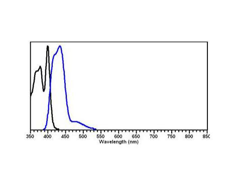
The emission spectra for this DyLight(TM) conjugate match the principle output wavelengths of most common fluorescence instrumentation. This product is designed for immunofluorescence microscopy, fluorescence based plate assays (FLISA) and fluorescent weste
Inhibition of 14-3-3 proteins leads to reduced numbers of synaptic GluN2B puncta in primary glutamatergic hippocampal neurons but not in cortical neurons.A, Representative images of YFP-difopein or YFP infected cortical neurons labeled with GluN2B (red), YFP (green), and SYP (blue) at DIV21.Bars, Illustrate numbers of synaptic GluN2B puncta/50 µm in YFP-difopein treated glutamatergic cortical neurons normalized to YFP controls (two-tailed unpaired Studentst-test: p = 0.6915, n = 8).B, Representative images of YFP-difopein or YFP infected hippocampal neurons labeled with GluN2B (red), YFP (green), and SYP (blue) at DIV21.Bars, Illustrate numbers of synaptic GluN2B puncta/50 µm in YFP-difopein treated glutamatergic hippocampal neurons normalized to YFP controls (two-tailed unpaired Studentst-test: *** p = 0.0005, n = 8-9). Error bars indicate SD. Scale bar, 10µm. Fig 3. PMID: 34962957.
Heterotopic neurons are born in mid- to late-gestation.A. Following injection of BrdU at E13.5, there are no BrdU+ neurons contained within PVNH (left panel) in a rat embryonically transfected withKiaa0319lshRNA on E15.5. There are transfected heterotopic neurons (red) (middle panel).B. Injection of BrdU at E15.5 labels large numbers of BrdU+ neurons (blue) within PVNH. Middle panel illustrate transfected neurons (green). Arrows indicate BrdU+ neurons that are co-labeled with eGFP (right panel).C. BrdU injected at E17.5 results in a population of BrdU+ neurons (blue) within PVNH, some of which are co-labeled (arrows). Wm = white matter. Bar = 200 µm in all panels. Goat Anti-Mouse IgG Fc DyLight(TM)405 (p/n 610-146-003) for BrdU staining. Fig 4. PMID: 23831424.
Inhibition of 14-3-3 proteins leads to reduced numbers of synaptic GluN2A puncta in primary glutamatergic cortical and hippocampal neurons. A, Representative images of YFP-difopein or YFP infected cortical neurons labeled with GluN2A (red), YFP (green), and SYP (blue) at DIV21.Bars, Illustrate numbers of synaptic GluN2A puncta/50 µm in YFP-difopein treated glutamatergic cortical neurons normalized to YFP controls (two-tailed unpaired Studentst-test: **** p<0.0001, n = 8).B, Representative images of YFP-difopein or YFP infected hippocampal neurons labeled with GluN2A (red), YFP (green), and SYP (blue) at DIV21.Bars, Illustrate numbers of synaptic GluN2A puncta/50 µm in YFP-difopein treated glutamatergic hippocampal neurons normalized to YFP controls (two-tailed unpaired Studentst-test: ** p = 0.0020, n = 9). Error bars indicate SD. Scale bar, 10µm. Fig 2. PMID: 34962957.
Inhibition of 14-3-3 proteins leads to reduced numbers of synaptic GluN1 puncta in primary glutamatergic cortical and hippocampal neurons.A, Representative images of YFP-difopein or YFP infected cortical neurons labeled with GluN1 (red), YFP (green), and SYP (blue) at DIV21.Bars, Illustrate numbers of synaptic GluN1 puncta/50 µm in YFP-difopein treated glutamatergic cortical neurons normalized to YFP controls (two-tailed unpaired Studentst-test: **** p<0.0001, n = 8).B, Representative images of YFP-difopein or YFP infected hippocampal neurons labeled with GluN1 (red), YFP (green), and SYP (blue) at DIV21.Bars, Illustrate numbers of synaptic GluN1 puncta/50 µm in YFP-difopein treated glutamatergic hippocampal neurons normalized to YFP controls (two-tailed unpaired Studentst-test: * p = 0.01413, n = 7). Error bars indicate SD. Scale bar, 10µm. Fig 1. PMID: 34962957.
Inhibition of 14-3-3 in the dCA1 increases the number of c-Fos-ir cells in the VTA during OFT. (B)Top, representative confocal images of c-Fos and TH co-staining revealing activation of neurons in the VTA of difopein- or YFP-injected mice following either 30-min OFT or gentle handling. Scale bar = 100 µm. Bottom, magnified representative image. Blue arrowheads point to examples of c-Fos and TH co-labeled cells (putative c-Fos-ir DA neurons). Orange arrowheads point to examples of c-Fos-ir only cells (putative c-Fos-ir non-DA neurons). S
* Mehrwertsteuer und Versandkosten nicht enthalten. Irrtümer und Preisänderungen vorbehalten