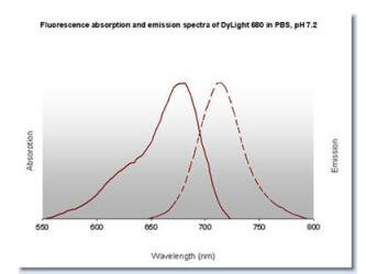
0.02 M Potassium Phosphate, 0.15 M Sodium Chloride, pH 7.2
Formulierung:
Lyophilized
Target-Kategorie:
Mouse
Antibody Type:
Secondary Antibody
Application Verdünnung:
FLISA: >1:20,000, IF Microscopy: >1:5,000, WB: >1:10,000
Anwendungsbeschreibung:
This product is designed for immunofluorescence microscopy, fluorescence based plate assays (FLISA) and fluorescent western blotting. This product is also suitable for multiplex analysis, including multicolor imaging, utilizing various commercial platfor
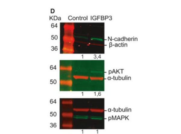
Western blot using Donkey Anti-Mouse IgG DyLight(TM)680.Insulin-like growth factor-binding protein 3 increases CRC cell migration and invasion through Akt activation. (D) Treatment with 50ngmL-1IGFBP-3 for 72h promotes the expression of N-cadherin in HCT116 cells as assessed by western blot (top panel). Phosphorylation status of Akt (middle panel) and MAPK (bottom panel) in HCT116 cells treated with 50ngmL-1IGFBP-3 for 15min. Representative images of three independent experiments (n=3). Numbers indicate the expression fold change relative to the loading control. Fig. 7. PMID: 32767843.
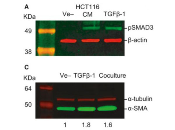
Western blot using Donkey Anti-Mouse IgG DyLight(TM)680. TGF-beta-mediated crosstalk between pericytes and CRC cells modulates pericyte secretome. (A) Incubation in HCT116 CM for 1h induces SMAD3 phosphorylation in PC, as assessed by western blot. Exogenous recombinant TGF-beta (10ngmL-1) was used as a positive control, and beta-actin was used as loading control (n=3). (C) Western blot showing increased expression of alphaSMA in PC cocultured with HCT116 cells or stimulated with 10 ngmL-1 TGF-beta1 for 48 h (n = 3). alpha-tubulin was used as loading control. Numbers indicate the expression fold change relative to the loading control. Fig. 5. PMID: 32767843.
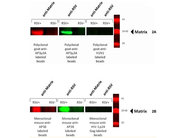
Western blot using Donkey Anti-Mouse IgG DyLight(TM)680.The HRSV M protein co-immunoprecipitates with the AP-3Mu3A and AP-3delta complex during HRSV infection. HEp2 cells at approximately 90% confluency were either infected at an MOI of 5 or mock infected for 24 hours, cells were scraped or proteins were subsequently extracted using MPER. Cell lysates were incubated for 6 hours with 1 µg of either polyclonal goat anti-AP-3Mu3A or monoclonal mouse anti-AP-3delta along with a antibody specific isotype control, polyclonal goat-anti H1N1or monoclonal mouse anti-HIV-1 p24 Gag at 4C on a rotating device. 20µl Protein A/G agarose beads were added to lysate plus corresponding antibody and incubated overnight. Immunoprecipate complex was pelleted and washed with PBS and then ran out on a SDS-PAGE gel and transferred to nitrocellulose membrane. Membrane was blocked and then probed with either monoclonal mouse anti-Matrix or polyclonal goat-anti HRSV primary antibody as described previously for one hour. Membranes were then washed with a PBS-Tween20 solution extensively and then probed with species-specific secondary antibodies donkey anti-goat DyLight800 and donkey anti-mouse DyLight680. Membranes were again washed extensively and blots were imaged on Odyssey Infrared imager. The results were reproducible in at least two independent assays. Fig 2. PMID: 29028839.
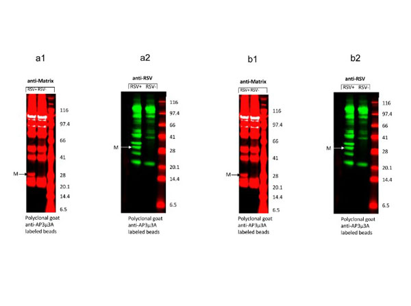
The HRSV M protein co-immunoprecipitates with the AP-3Mu3A complex during HRSV infection. a1)The HRSV M protein co-immunoprecipitates with the AP-3Mu3A complex during HRSV infection. HEp2 cells at approximately 90% confluency were either infected at an MOI of 5 or mock infected for 24 hours, cells were scraped or proteins were subsequently extracted using MPER. Cell lysates were incubated for 6 hours with 1 µg of polyclonal goat anti-AP-3Mu3A at 4C on a rotating device. 20µl Protein A/G agarose beads were added to lysate plus corresponding antibody and incubated overnight. Immunoprecipate complex was pelleted and washed with PBS and then ran out on a SDS-PAGE gel and transferred to nitrocellulose membrane. Membrane was blocked and then probed with monoclonal mouse anti-Matrix primary antibody as described previously for one hour. Membranes were then washed with a PBS-Tween20 solution extensively and then probed with species-specific secondary antibodies donkey anti-mouse DyLight(TM)680. Membranes were again washed extensively and blots were imaged on Odyssey Infrared imager. The last lane shows protein molecular weight marker (KDa). The results were reproducible in at least two independent assays.a2)The HRSV M protein co-immunoprecipitates with the AP-3Mu3A complex during HRSV infection. HEp2 cells at approximately 90% confluency were either infe
* Mehrwertsteuer und Versandkosten nicht enthalten. Irrtümer und Preisänderungen vorbehalten