Mouse Anti-Rabbit IgG Biotin Conjugate has been assayed against 1.0 ug of Rabbit IgG in a standard capture ELISA using Peroxidase Conjugated Streptavidin S000-03 and ABTS (2,2-azino-bis-[3-ethylbenthiazoline-6-sulfonic acid]) code ABTS-100 as a subst
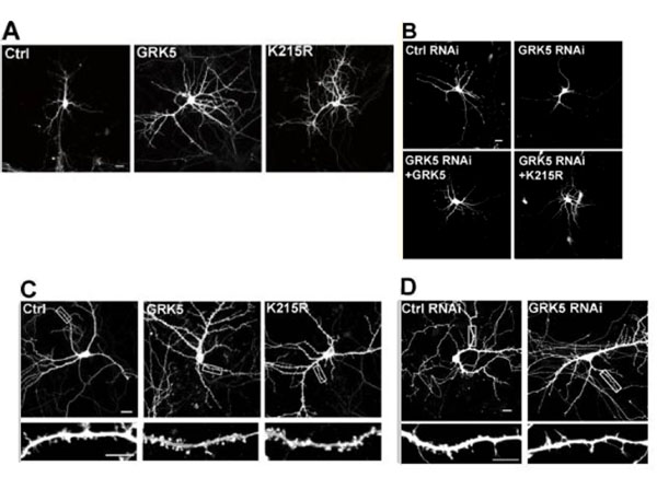
GRK5 regulates dendritic development.(A and B) Hippocampal neuron cultures transfected at DIV5 were observed at DIV8. Total dendritic branch tip numbers (TDBTN) and total dendrite length of transfected neurons were measured. For each group, 40-60 (A) or 30-40 (B) neurons from three independent cultures were analyzed. One-way ANOVA followed by Tukey-Kramer posthoc test. (C and D) Hippocampal neurons were transfected at DIV9 and observed at DIV17. Boxed regions are enlarged below each image. For each group, 30-40 dendrites of 8-10 neurons from three independent cultures were analyzed. Protrusion and spine number were measured. (C) GFP was cotransfected with GRK5 variants to visualize dendritic spines (one-way ANOVA followed by Tukey-Kramer posthoc test). (D) Neuron cultures transfected with control or GRK5 RNAi constructs (Studentsttest). Bars, 10 µm. Error bars indicate SEM. *, P < 0.03, **, P < 0.01, ***, P < 0.001. Ctrl, control. Figure 1. PMID: 21930777.
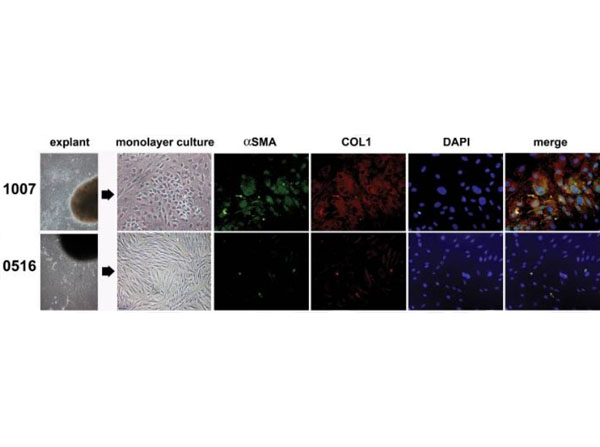
Mouse Anti-Rabbit IgG biotin conjugated antibody.Peri-Urethral Prostate Tissues Exhibit Fibroblastic and Myofibroblastic Cell Populations. Peri-urethral prostate tissues from patients 1007 and 0516 were explanted and primary fibroblasts were isolated and grown to monolayer cultures. Photomicrographs demonstrate fibroblastic morphology for 0516 primary cells but mixed fibroblastic and myofibroblastic morphologies for patient 1007. Cells from both cultures were then stained for collagen 1 (COL1) (PE-cy5-conjugated Ab, red), alpha-smooth muscle actin (alphaSMA) (fluorescein-conjugated Ab, green), or the nuclei counterstained with DAPI (blue). Merged images show that primary cells from patient 1007 exhibited high levels of co-localized COL1 and alphaSMA protein expression (yellow) consistent with a myofibroblastic phenotype. Control mouse IgG2a and rabbit IgG biotin conjugate (p/n 611-306-122) were used at 1∶2000 dilution. All images were captured at 400X in visible light on brightfield settings. Figure 1. PMID: 23173053.
* Mehrwertsteuer und Versandkosten nicht enthalten. Irrtümer und Preisänderungen vorbehalten