
Rabbit IgG (H&L) Antibody DyLight(TM) 800 Conjugated Pre-Adsorbed, DL800, Sheep, Polyclonal
Artikelnummer:
ROC-611-645-122
- Bilder (9)

| Artikelname: | Rabbit IgG (H&L) Antibody DyLight(TM) 800 Conjugated Pre-Adsorbed, DL800, Sheep, Polyclonal |
| Artikelnummer: | ROC-611-645-122 |
| Hersteller Artikelnummer: | 611-645-122 |
| Alternativnummer: | ROC-611-645-122 |
| Hersteller: | Rockland Immunochemicals |
| Wirt: | Sheep |
| Kategorie: | Antikörper |
| Spezies Reaktivität: | Rabbit |
| Immunogen: | Rabbit IgG whole molecule |
| Konjugation: | DL800 |
| Alternative Synonym: | Sheep Anti Rabbit IgG Antibody DyLight 800(TM) Conjugate, Sheep Anti-Rabbit IgG DyLight 800(TM) Conjugated Antibody |
| Klonalität: | Polyclonal |
| Konzentration: | 1.0 mg/mL by UV absorbance at 280 nm |
| Puffer: | 0.02 M Potassium Phosphate, 0.15 M Sodium Chloride, pH 7.2 |
| Formulierung: | Lyophilized |
| Target-Kategorie: | Rabbit |
| Antibody Type: | Secondary Antibody |
| Application Verdünnung: | FLISA: >1:20,000, IF Microscopy: >1:5,000, WB: >1:10,000 |
| Anwendungsbeschreibung: | This product is designed for immunofluorescence microscopy, fluorescence based plate assays (FLISA) and fluorescent western blotting. This product is also suitable for multiplex analysis, including multicolor imaging, utilizing various commercial platfor |
|
|
|
|
|
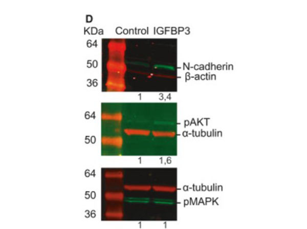
Western Blot results using Sheep Anti-Rabbit IgG DyLight(TM)800.Insulin-like growth factor-binding protein 3 increases CRC cell migration and invasion through Akt activation. (D) Treatment with 50ngmL-1IGFBP-3 for 72h promotes the expression of N-cadherin in HCT116 cells as assessed by western blot (top panel). Phosphorylation status of Akt (middle panel) and MAPK (bottom panel) in HCT116 cells treated with 50ngmL-1IGFBP-3 for 15min. Representative images of three independent experiments (n=3). Numbers indicate the expression fold change relative to the loading control. Fig. 7. PMID: 32767843. |
|
|
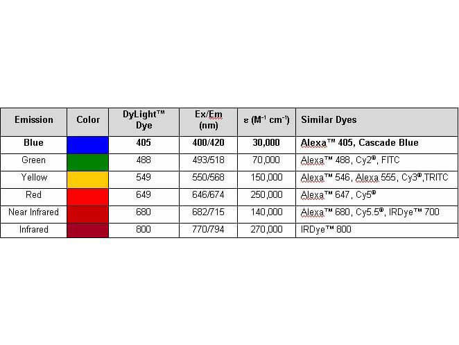
Properties of DyLight(TM) Conjugates. |
|
|
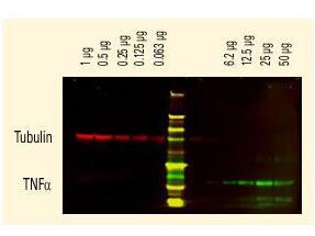
DyLight(TM) dyes can be used for two-color western blot detection with low background and high signal. Anti-tubulin was detected using a DyLight(TM) 680 conjugate. Anti-TNFa was detected using a DyLight(TM) 800 conjugate. The image was captured using the Odyssey Infrared Imaging System developed by LI-COR. |
|
|
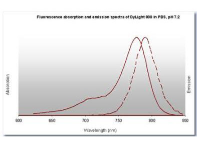
DyLight(TM) 800 Fluorescence Spectra. |
|
|
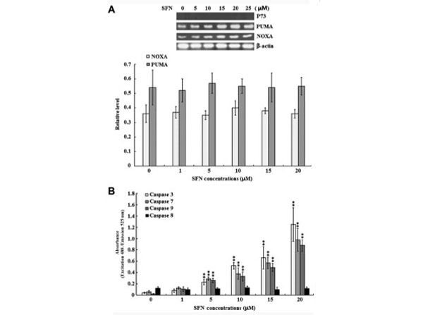
Western Blot results using Sheep Anti-Rabbit IgG DyLight(TM)800.Effect of SFN on p73 expression and caspase enzyme activities. SW480 cells were treated with 0, 1, 5, 10, 15 and 20 µM SFN for 24 h. (A) The protein expression levels of p73, PUMA and NOXA were examined by western blotting. Quantified band densities are summarized below the images of the bands, and target protein expression levels were normalized to that of b-actin. (B) The activity of caspase-3, -7, -8 and -9 was determined using specific ELISA kits. The results of three independent experiments are shown. **P<0.01 vs. 0 µM SFN. SFN, sulforaphane, PUMA, p53 upregulated modulator of apoptosis, NOXA, phorbol-12-myristate-13-acetate-induced protein 1, ELISA, enzyme-linked immunosorbent assay. Figure2. PMID: 28944886. |
|
|
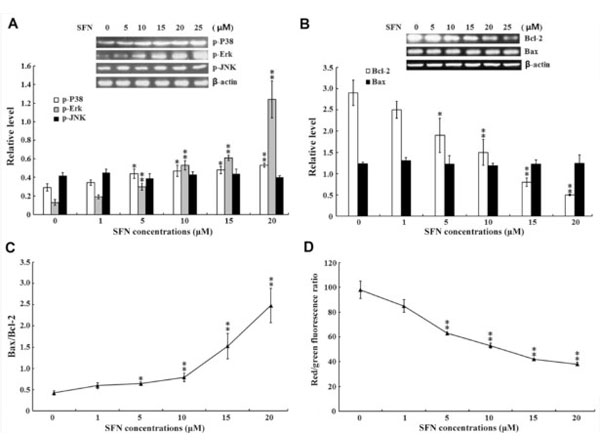
Western Blot results using Sheep Anti-Rabbit IgG DyLight(TM)800.Effect of SFN on MAPKs and intrinsic apoptotic signaling pathways. SW480 cells were treated with 0, 1, 5, 10, 15 and 20 µM SFN for 24 h. (A) The protein expression levels of (A) p-p38, p-Erk and p-JNK and (B) Bcl-2 and Bax were examined by western blotting, and the quantified results are summarized below the images of the protein bands. Target protein expression levels were normalized to that of b-actin. (C) The Bax/Bcl-2 ratio. (D) The MMP was examined using a MMP assay kit with a JC-1 probe. The results of three independent experiments are shown. *P<0.05 and **P<0.01 vs. 0 µM SFN. SFN, sulforaphane, MAPK, mitogen-activated protein kinases, p-, phosphorylated, Erk, extracellular signal-regulated kinases, JNK, c-Jun N-terminal kinases, Bcl-2, B-cell lymphoma-2, Bax, Bcl-2-associated protein X, MMP, mitochondrial membrane potential. Figure3. PMID: 28944886. |
|
|
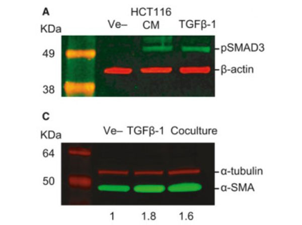
Western Blot using Sheep Anti-Rabbit IgG DyLight(TM)800.TGF-beta-mediated crosstalk between pericytes and CRC cells modulates pericyte secretome. (A) Incubation in HCT116 CM for 1h induces SMAD3 phosphorylation in PC, as assessed by western blot. Exogenous recombinant TGF-beta (10ngmL-1) was used as a positive control, and beta-actin was used as loading control (n=3). (C) Western blot showing increased expression of alphaSMA in PC cocultured with HCT116 cells or stimulated with 10ngmL-1TGF-beta1 for 48h (n=3). alpha-tubulin was used as loading control. Numbers indicate the expression fold change relative to the loading control. Fig. 5. PMID: 32767843. |
|
|
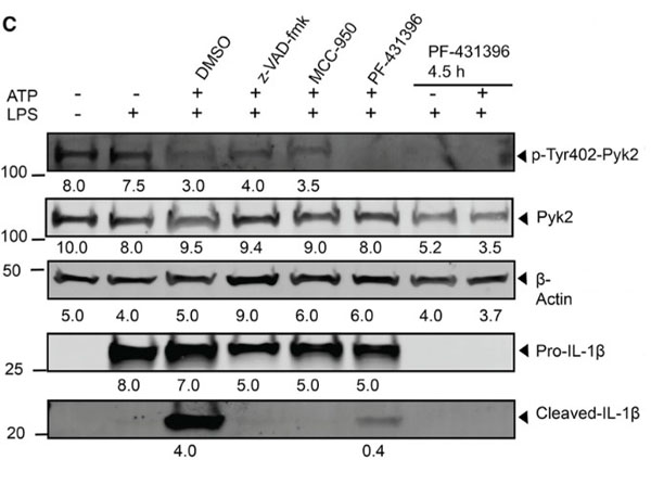
Western Blot Results using Sheep Anti-Rabbit IgG DyLight(TM)800.LPL stabilizes Pyk2-ASC interaction by regulating Pyk2 localization.(C) WT BMDMs were primed with LPS and stimulated for NLRP3 activation by ATP (30 min) in the absence and presence of z-VAD-fmk (50 µM, 1 h), MCC-950 (50 µM, 1 h), and PF-431396 (25 µM) for 1 h or 4.5 h. Cell lysates were probed for Pyk2 phosphorylation and IL-1beta products by immunoblotting. The density of each band is shown below. Here, beta-actin is the total cell control. Figure 4. PMID: 35294888. |
Produktgarantie und fachkundiger Support
