![]()
|
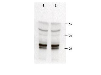
Rabbit anti-cdk2 (100-401-161) was used at a 1:200 dilution to detect cdk-2 in asynchronous HeLa cell lysates (run in duplicate). Approximately 50 µg of lysate was separated on a 15% SDS-PAGE gel. Cdk-2 is indicated by an arrowhead as a 32-33 kDa band. Note that multiple isoforms as well as phosphorylated forms of cdk-2 may be detected by this antibody. Primary antibody was reacted at room temperature for 1 h. After subsequent washing, a 1:5,000 dilution of HRP conjugated Gt-a-Rabbit IgG (611-103-122) preceded color development. |
![]()
|
|
![]()
|
This product is assembled as a kit. See attached protocol or CofA for further details. |
![]()
|
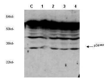
Rocklands anti-cdc2 Cyclin Dependent Kinase (100-401-160) was used to detect human p34cdc2by western blot in untreated (Contol) and drug treated lysates of MCF-7 cells. Lane 1-4 represents 3.1?uM, 6.2?uM, 12.5?uM and 25.0?uM genistein treatment of cells before lysates were prepared. Detection occurs using a 1:1,000 dilution. Although this antiserum detects non-specific bands at higher MW, a clear induction of signal is observed as the concentration of drug is increased. Personnel Communication, Xiao He Yang, University of Oklahoma Health Sciences Center. |
![]()
|
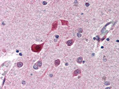
Rocklands anti-CDK5 antibody (100-401-163) was diluted 1:500 to detect CDK5 in human brain cortex tissue. Tissue was formalin fixed and paraffin embedded. No pre-treatment of sample was required. The image shows the localization of antibody as the precipitated red signal, with a hematoxylin purple nuclear counter stain. |
![]()
|
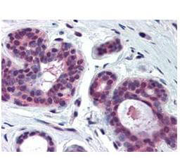
Rocklands Anti-CDK2 antibody (100-401-161) was diluted 1:500 to detect CDK2 in human skin tissue. Tissue was formalin fixed and paraffin embedded. No pre-treatment of sample was required. The image shows the localization of antibody as the precipitated red signal, with a hematoxylin purple nuclear counter stain. |
![]()
|
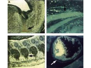
Immunocytochemical staining of mouse tissue using anti-cdk9 (PITALRE) antiserum (100-401-167). The staining shows the location of mcdk9/PITALRE protein in developing mouse tissue. Arrows indicate areas of high expression. Panel A: Peroxidase-DAB immunostaining of mcdk9/PITALRE protein in the developing mouse brain in the differentiated region of the medulla oblongata just below the fourth ventricle. Similar staining is shown in Panel B in the dorsal root ganglia. Panel C: Fluorescein immunofluorescence of mcdk9IPITALRE in skeletal muscle. Similar staining is shown in Panel D in cardiac muscle. Other detection systems should yield similar results. Sections from each specimen were cut at 5-7 µm, mounted on glass and dried overnight at 37C. All sections then were deparaffinized in xylene, rehydrated through a graded alcohol series and washed in phosphate-buffered saline (PBS). PBS was used for all subsequent washes and for antiserum dilution. Tissue sections were quenched sequentially in 0.5% hydrogen peroxide and blocked with diluted 10% normal goat anti-rabbit serum. Slides were incubated at 20 C for 1 h with rabbit anti-cdk9 (1:500) dilution, washed, and then reacted with diluted goat anti-rabbit biotinylated antibody for 30 min. All the slides were then reacted with streptavidin-peroxidase conjugate for 30 min at 20 C. Diaminobenzidine was used as the final chromogen and hematoxylin was used as the nuclear counterstain. Negative controls for each tissue section were prepared by substituting the primary antiserum with pre-immune serum. |