
Streptavidin Peroxidase Conjugated, HRP
Artikelnummer:
ROC-S000-03
- Bilder (11)
| Artikelname: | Streptavidin Peroxidase Conjugated, HRP |
| Artikelnummer: | ROC-S000-03 |
| Hersteller Artikelnummer: | S000-03 |
| Alternativnummer: | ROC-S000-03 |
| Hersteller: | Rockland Immunochemicals |
| Kategorie: | Sonstiges |
| Applikation: | DOT, WB |
| Konjugation: | HRP |
| Alternative Synonym: | HRP-SA, Horseradish Peroxidase conjugated S avidin, Streptavidin HRP, Streptavidin conjugated to horseradish peroxidase (HRP), HRP-linked Streptavidin |
| Application Verdünnung: | ELISA: 1:20,000 - 1:200,000, IHC: 1:1,000 - 1:5,000, IF Microscopy: User Optimized, WB: 1:10,000 - 1:40,000 |
| Anwendungsbeschreibung: | Streptavidin Peroxidase has been tested by dot blot and western blot and is a useful detection reagent for primary antibodies conjugated to biotin. Streptavidin Peroxidase can be utilized in Immunohistochemistry, Immunofluorescence, immuno-EM, Western Bl |
|
|
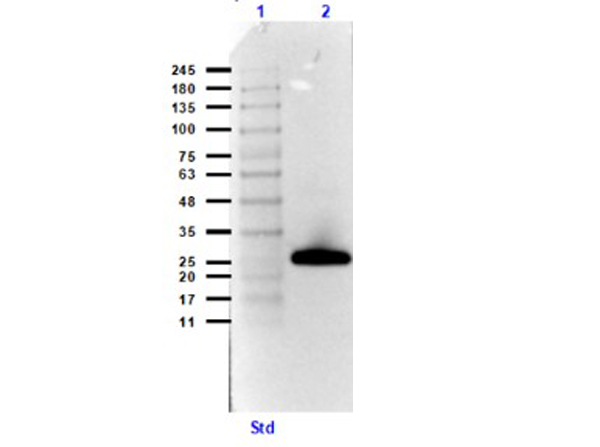
Western Blot Results using Streptavidin Peroxidase Conjugate and Goat Anti-GST Biotin Conjugate Antibodies. Lane 1: Opal prestained molecular weight ladder (p/n MB-210-0500). Lane 2: GST (p/n 000-001-200) [0.05 µg]. Primary Antibody: Goat Anti-GST Biotin Conjugate (p/n 600-106-200) at 1.0µg/mL overnight at 4C. Secondary Antibody, Streptavidin Peroxidase Conjugate at 1:40,000 for 30mins at RT.Block: Blocking Buffer for Fluorescent Western Blotting (p/n MB-070) for 30mins at RT. Exp: 5 sec. |
|
|
Streptavidin Peroxidase Conjugated |
|
|
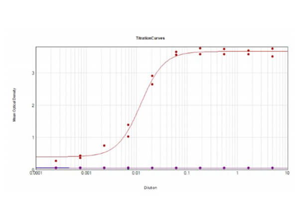
ELISA Results of Human IgG Whole Molecule Biotin Conjugated using Streptavidin-HRP. Each well was coated in duplicate with 1.0 µg of Human IgG Whole Molecule Biotin Conjugate. The working dilution is 82,800. The starting dilution of antibody was 5µg/ml and the X-axis represents the Log10 of a 3-fold dilution. This titration is a 4-parameter curve fit where the IC50 is defined as the titer of the antibody. Assay performed using Streptavidin-HRP (p/n S000-03) and TMB substrate (p/n TMBE-1000). |
|
|
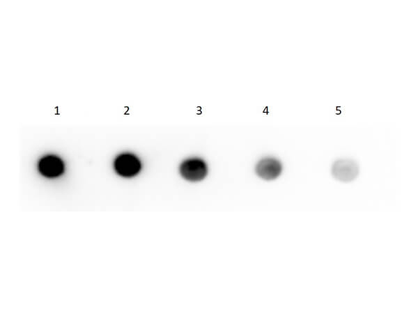
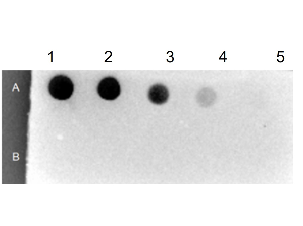
Dot Blot of Human IgG F(c) Fragment Biotin Conjugated using Streptavidin HRP. Human IgG F(c) Biotin Conjugate (1) 100ng, (2) 33.33ng, (3) 11.11ng, (4) 3.70ng, (5) 1.23ng. Primary Antibody: none. Secondary Antibody: Streptavidin HRP (p/n S000-03) at 1:40,000 for 30 mins at RT. Block: BlockOut buffer (p/n MB-073) at RT for 30 mins. Exposure: 1 sec. |
|
|
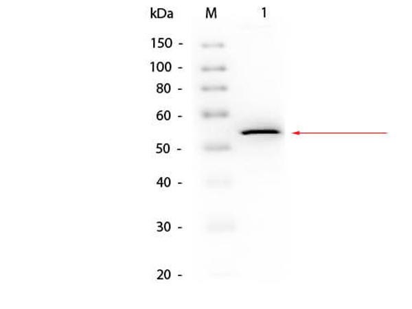
Western Blot of Goat anti-Glycerol Kinase Antibody Biotin Conjugated using Streptavidin HRP. Lane 1: Glycerol Kinase. Load: 50 ng per lane. Primary antibody: Glycerol Kinase Antibody Biotin Conjugated at 1:1000 overnight at 4C. Secondary antibody: HRP Streptavidin (p/n S000-03) secondary antibody at 1:40,000 for 30 min at RT. Block: MB-070 for 30 min at RT. Predicted/Observed size: 55 kDa, 55 kDa for Glycerol Kinase. |
|
|
Dot Blot Results of Streptavidin Peroxidase Conjugate. Row A: BSA-Biotin Conjugated. Row B: BSA. Sample dilutions: 1- 100ng, 2- 33.33ng, 3- 11.11ng, 4-3.7ng, 5- 1.23ng. Streptavidin Peroxidase Conjugated at 1.0µg/mL for 30mins at RT. |
|
|
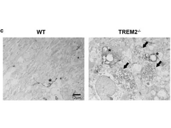
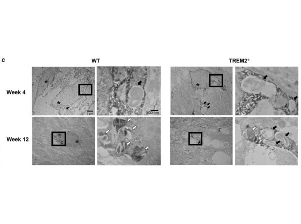
Immuno-electron microscopy using biotinylated anti-rabbit and streptavidin-HRP.TREM2-/-mice show more severe axonal pathology after CPZ |
|
|
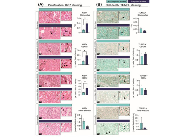
Percentage of cells showing cell-cycle activation as informed by Ki67 immunostaining (A) and cell death as identified by TUNEL assay (B) in the kidney in response to pregnancy in mice.Representative image of stained kidney from non-pregnant and pregnant female mice. Data are presented as mean SEM (n= 5/group). Asterisks represent significant differences between non-pregnant and pregnant mice as determined by Studentst-Test (*p< 0.05). Slides incubated with goat anti-rabbit secondary antibody (1:1000) and streptavidin-horseradish peroxidase (1:500,p/n S000-03). Images with the labels i and ii depict high magnification of the selected area. Arrow heads indicate positive DAB staining. Scale bar is 50 µm. Figure 3. PMID: 35682969. |
|
|
|
|
|
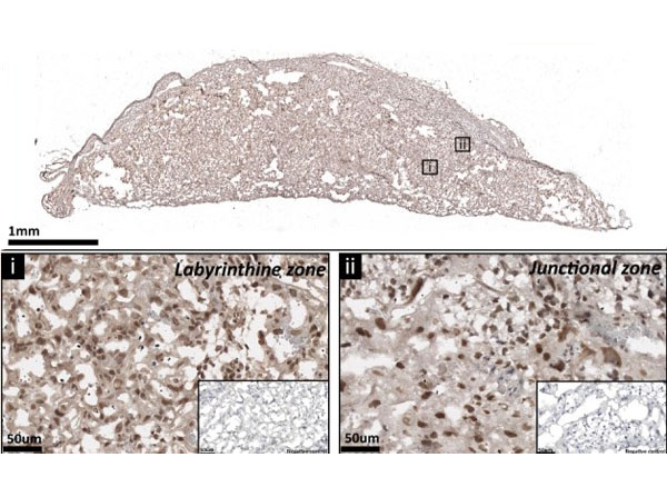
The expression of p110alpha protein by the mouse placenta on day 19 of pregnancy. Representative stained section shown with negative control shown in the figure inset. For localization of p110alpha, placental sections were washed with PBS to remove OCT and underwent antigen retrieval with citrate buffer before immunolabelling against p110alpha. Sections were treated with 0.5% Triton X-100 before immunolabelling. Bound antibody was detected using biotinylated goat anti-rabbit IgG followed by streptavidin-conjugated horseradish peroxidase (p/n S000-03) and 3,3-diaminobenzidine (DAB). Sections were lightly counterstained with hematoxylin and mounted in DPX. Supplement Fig 1. PMID: 31241463. |
|
|
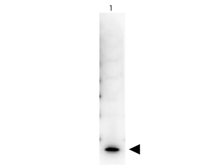
Western Blot of Peroxidase Conjugated Streptavidin. Lane 1: Human IL-7. Load: 50 ng per lane. Primary antibody: Human IL-7 Biotin Conjugated antibody at 1:1,000 for overnight at 4C. Secondary antibody: Peroxidase Conjugated Streptavidin at 1:40,000 for 30 min at RT. Block: 5% BLOTTO 30 min at RT. Predicted/Observed size: 17 kDa, 17 kDa for Human IL-7. Other band(s): none. |
Produktgarantie und fachkundiger Support
