SA, S avidin, streptococcus avidin, streptavidin DyLight(TM) 488 Conjugated
Konzentration:
1 mg/mL by UV absorbance at 280 nm
Puffer:
0.02 M Potassium Phosphate, 0.15 M Sodium Chloride, pH 7.2
Formulierung:
Lyophilized
Application Verdünnung:
FLISA: >1:20,000, IF Microscopy: >1:5,000, WB: >1:10,000
Anwendungsbeschreibung:
Streptavidin DyLight(TM)488 has been tested by western blot and immunofluorescence and is designed for immunofluorescence microscopy, fluorescence based plate assays (FLISA) and fluorescent western blotting. This product is also suitable for multiplex analy
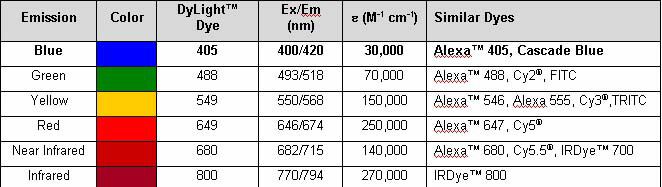
Properties of DyLight(TM) Fluorescent Dyes.
Streptavidin DyLight(TM) 488 Conjugated
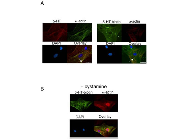
5-HT and 5-HT-biotin localize to alpha-actin and are incorporated into proteins. A. Immunocytochemistry of aortic smooth muscle cells incubated with exogenous 5-HT (12.7 µM, left) or 5-HT biotin (12.7 µM, right) and alpha-actin for 1 hour prior to fixation and visualization using an anti-rabbit fluorescent secondary (for 5-HT) or DyLight(TM) 488 streptavidin (p/n S000-41) secondary (for 5-HT biotin). Representative of four different aortic explants. B. Effect of cystamine (10 mM) on 5-HT-biotin localization in aortic smooth muscle cells. Representative of four different aortic explants. Fig 6. PMID: 19479059.
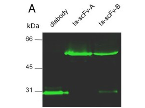
Characterization of secreted bispecific antibodies. (A) Engineered, bispecific antibodies secreted into the conditioned medium of stably transfected HEK293 cells (293diabody, 293ta-scFv-Aor 293ta-scFv-B) were characterized for expression levels and binding properties. (A) western blot analysis with DyLight 488 conjugated-streptavidin (p/n S000-41). Migration distances of molecular mass markers are indicated (kDa). The blot was developed with anti-His tag mAb. Figure2.PMID: 25057445.
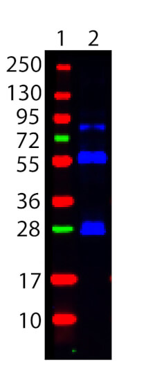
Western Blot showing detection Biotin. 100 ng of Biotin conjugated Guinea Pig IgG (Lane 2) was run on a 4-20% gel and transferred to 0.45 µm nitrocellulose. After blocking with 1% BSA-TTBS (p/n MB-013, diluted to 1X) 30 min at 20C, STREPTAVIDIN DyLight(TM) 488 Conjugated (p/n S000-41) was used at 1:1000 in Blocking Buffer for Fluorescent Western Blotting (p/n MB-070) and imaged using the Bio-Rad VersaDoc 4000 MP. Molecular weight markers are in lane 1.
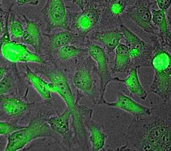
Rockland Dylight 488 conjugated Streptavidin was used to stain HeLa cells by immunofluorescence. HeLa cells were plated in 12 well plates, fixed for 5 min in 1:1 MeTOH:Acetone, blocked with MB-071 (preservative free) for 15 min and stained 1 hr with Rockland biotin conjugated anti lactate dehydrogenase antibody (p/n 200-1673 lot 5412 1:200 in blocking buffer). Plate was washed 3X in PBS, and primary biotin conjugate was detected using DyLight 488 conjugated Streptavidin (S000-41 lot 21097) 1:10000 for 30 min. Well was washed 3X in PBS. Image was taken using EVOS fl All Digital Inverted Fluorescence Microscope by AMG (Advanced Microscopy Group).
* Mehrwertsteuer und Versandkosten nicht enthalten. Irrtümer und Preisänderungen vorbehalten