
ExoQuick-CG cGMP-grade Exosome Isolation reagent ()
- Bilder (2)


| Artikelname: | ExoQuick-CG cGMP-grade Exosome Isolation reagent () |
| Artikelnummer: | SBI-EXOCG50A-1 |
| Hersteller Artikelnummer: | EXOCG50A-1 |
| Alternativnummer: | SBI-EXOCG50A-1 |
| Hersteller: | System Biosciences |
| Kategorie: | Molekularbiologie |
| ExoQuick-CG cGMP-grade Exosome Isolation reagent (50 ml) |
ExoQuick-CGExoQuick is now ready for purifying exosomes for clinical use with a version of ExoQuick manufactured in compliance with 21 CFR 820. ExoQuick-CG: cGMP quality exosome isolation for pre-clinical applications "We therefore pursued the ExoQuick® method for further study, as these samples required much less sample input, a key benefit when working with clinical samples and mouse models1." Based on SBI’s well-regarded ExoQuick-TC, SBI's ExoQuick-CG exosome precipitation reagent makes miRNA and protein biomarker discoveries simple, reliable, and quantitative. Enrich for exosomal miRNAs with ExoQuick-CG and accurately profile them using SBI's SeraMir qPCR arrays. Downstream protein analysis is also possible with SBI's exosome specific antibodies and ELISA kits. Enjoy the same benefits of ExoQuick-TC as well as these additional benefits:
Like ExoQuick-TC, ExoQuick-CG enables high-throughput, quantitative isolation of exosomes from most biofluids. Compatible with a wide variety of downstream applications, ExoQuick-CG is an effective and proven alternative to ultracentrifugation1-3.
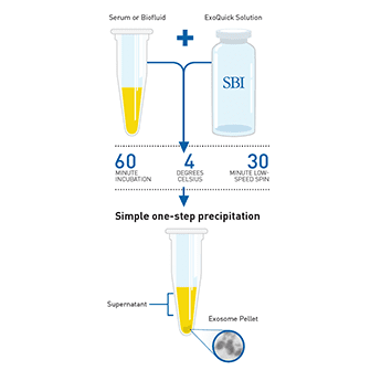
How It WorksHigh-throughput, quantitative exosome recovery Just as with ExoQuick-TC, ExoQuick-CG can be used to purify exosomes from saliva1, urine2, follicular fluid3, tissue culture media4, and breast milk5. With a simple workflow involving minimal hands-on time and low input sample volume requirements, ExoQuick-CG is an excellent option for researchers who need to purify exosomes for pre-clinical, in vivo studies. To isolate exosomes from cleared serum, plasma, or ascites fluid, simply:
Isolated exosomes can be found in the pellet and resuspended in an appropriate solution.
You can verify the presence of exosomes with a number of different methods, including Western blotting for general exosome markers (CD63, CD9, CD81, and HSP70), NanoSight analysis, or EM (learn about different ways to detect exosomes and more in our Exosome Basics Guide). The Bottom Line
Supporting DataExosome Size Distribution and Yields Using ExoQuick-CG
Figure 1. 3.3 mL of ExoQuick-CG was combined with 10 mL of conditioned media from either Human HT1080 lung sarcoma cells or Mouse JAWS II dendritic cells that had been cultured in Exo-FBS (exosome depleted FBS medium supplement, catalog # EXO-FBS-50A-1). All samples were incubated overnight at 4°C for exosome precipitation. Exosomes were resuspended in 0.5 mL of PBS and visualized on a NanoSight LM10 instrument.
Western blotting of the ExoQuick-CG-isolated particles using an exosome-specific antibody (anti-CD63) further validates that the particles isolated with ExoQuick-CG are exosomes (Figure 2). Figure 2. Approximately 10-50 µg of exosomes from the NanoSight studies shown in Figure 1 were separated via denaturing SDS PAGE for Western blot analysis. CD63 protein was detected using SBI’s rabbit anti-CD63 primary antibody and SBI’s HRP-conjugated secondary goat anti-rabbit antibody (catalog # EXOAB-CD63A-1). |
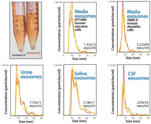 NanoSight analysis shows ExoQuick-CG isolates high concentrations of exosomes from a variety of biofluids, and that the isolated exosomes are in the expected size range (Figure 1).
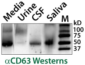 Exosome CD63 Western Blot Validation Studies
| Lagerung: | See Manual |
|
|
Figure 1. 3.3 mL of ExoQuick-CG was combined with 10 mL of conditioned media from either Human HT1080 lung sarcoma cells or Mouse JAWS II dendritic cells that had been cultured in Exo-FBS (exosome depleted FBS medium supplement, catalog # EXO-FBS-50A-1). All samples were incubated overnight at 4°C for exosome precipitation. Exosomes were resuspended in 0.5 mL of PBS and visualized on a NanoSight LM10 instrument. |
|
|
Figure 2. Approximately 10-50 µg of exosomes from the NanoSight studies shown in Figure 1 were separated via denaturing SDS PAGE for Western blot analysis. CD63 protein was detected using SBI’s rabbit anti-CD63 primary antibody and SBI’s HRP-conjugated secondary goat anti-rabbit antibody (catalog # EXOAB-CD63A-1). |
