
STIP1 Rabbit mAb, Unconjugated
Artikelnummer:
ABB-A0036
- Bilder (6)


| Artikelname: | STIP1 Rabbit mAb, Unconjugated |
| Artikelnummer: | ABB-A0036 |
| Hersteller Artikelnummer: | A0036 |
| Alternativnummer: | ABB-A0036-20UL, ABB-A0036-100UL |
| Hersteller: | ABclonal |
| Wirt: | Rabbit |
| Kategorie: | Antikörper |
| Applikation: | ELISA, IHC-P, WB |
| Spezies Reaktivität: | Human |
| Immunogen: | Recombinant protein (or fragment).This information is considered to be commercially sensitive. |
| Konjugation: | Unconjugated |
| Alternative Synonym: | HOP, P60, STI1, STI1L, HEL-S-94n, IEF-SSP-3521, STIP1 |
| STIP1 is an adaptor protein that coordinates the functions of HSP70 (see HSPA1A, MIM 140550) and HSP90 (see HSP90AA1, MIM 140571) in protein folding. It is thought to assist in the transfer of proteins from HSP70 to HSP90 by binding both HSP90 and substrate-bound HSP70. STIP1 also stimulates the ATPase activity of HSP70 and inhibits the ATPase activity of HSP90, suggesting that it regulates both the conformations and ATPase cycles of these chaperones (Song and Masison, 2005 [PubMed 16100115]). |
| Application Verdünnung: | WB,1:500 - 1:2000|IHC-P,1:50 - 1:200|ELISA,Recommended starting concentration is 1 µg/mL. Please optimize the concentration based on your specific assay requirements. |
| Anwendungsbeschreibung: | Cross-Reactivity: Human,Mouse,Rat, ResearchArea: Epigenetics Nuclear Signaling,RNA Binding,Signal Transduction,Neuroscience,Neurodegenerative Diseases. |
|
|
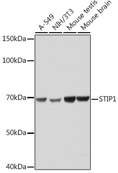
Western blot analysis of various lysates using STIP1 Rabbit mAb (A0036) at 1:1000 dilution. Secondary antibody: HRP-conjugated Goat anti-Rabbit IgG (H+L) (AS014) at 1:10000 dilution. Lysates/proteins: 25µg per lane. Blocking buffer: 3% nonfat dry milk in TBST. Detection: ECL Basic Kit (RM00020). Exposure time: 1s. |
|
|
Western blot analysis of various lysates using STIP1 Rabbit mAb (A0036) at 1:1000 dilution. Secondary antibody: HRP-conjugated Goat anti-Rabbit IgG (H+L) (AS014) at 1:10000 dilution. Lysates/proteins: 25µg per lane. Blocking buffer: 3% nonfat dry milk in TBST. Detection: ECL Basic Kit (RM00020). Exposure time: 10s. |
|
|
Immunohistochemistry analysis of paraffin-embedded Human lung adenocarcinoma tissue using STIP1 Rabbit mAb (A0036) at a dilution of 1:200 (40x lens). High pressure antigen retrieval performed with 0.01M Citrate buffer (pH 6.0) prior to IHC staining. |
|
|
Immunohistochemistry analysis of paraffin-embedded Mouse brain tissue using STIP1 Rabbit mAb (A0036) at a dilution of 1:200 (40x lens). High pressure antigen retrieval performed with 0.01M Citrate buffer (pH 6.0) prior to IHC staining. |
|
|
Immunohistochemistry analysis of paraffin-embedded Mouse testis tissue using STIP1 Rabbit mAb (A0036) at a dilution of 1:200 (40x lens). High pressure antigen retrieval performed with 0.01M Citrate buffer (pH 6.0) prior to IHC staining. |
|
|
Immunohistochemistry analysis of paraffin-embedded Rat lung tissue using STIP1 Rabbit mAb (A0036) at a dilution of 1:200 (40x lens). High pressure antigen retrieval performed with 0.01M Citrate buffer (pH 6.0) prior to IHC staining. |
Produktgarantie und fachkundiger Support
