
PGP9.5/UCHL1 Rabbit mAb, Unconjugated, Monoclonal
Artikelnummer:
ABB-A20380
- Bilder (9)

| Artikelname: | PGP9.5/UCHL1 Rabbit mAb, Unconjugated, Monoclonal |
| Artikelnummer: | ABB-A20380 |
| Hersteller Artikelnummer: | A20380 |
| Alternativnummer: | ABB-A20380-100UL,ABB-A20380-20UL |
| Hersteller: | ABclonal |
| Wirt: | Rabbit |
| Kategorie: | Antikörper |
| Applikation: | ELISA, IF, IHC-P, WB |
| Spezies Reaktivität: | Human |
| Immunogen: | Recombinant protein (or fragment).This information is considered to be commercially sensitive. |
| Konjugation: | Unconjugated |
| Alternative Synonym: | NDGOA, PARK5, PGP95, SPG79, PGP9.5, SPG79A, UCHL-1, Uch-L1, HEL-117, PGP 9.5, HEL-S-53, PGP9.5/UCHL1 |
| The protein encoded by this gene belongs to the peptidase C12 family. This enzyme is a thiol protease that hydrolyzes a peptide bond at the C-terminal glycine of ubiquitin. This gene is specifically expressed in the neurons and in cells of the diffuse neuroendocrine system. Mutations in this gene may be associated with Parkinson disease. |
| Application Verdünnung: | WB,1:10000 - 1:50000|IF/ICC,1:500 - 1:1000|IF-P,1:500 - 1:1000|IHC-P,1:500 - 1:1000|ELISA,Recommended starting concentration is 1 µg/mL. Please optimize the concentration based on your specific assay requirements. |
| Anwendungsbeschreibung: | Cross-Reactivity: Human,Mouse,Rat. ResearchArea: Cell Biology Developmental Biology,Ubiquitin,Ubiquitin-Proteasome Signaling Pathway,Neuroscience, Cell Type Marker,Neurodegenerative Diseases,Dopamine Signaling in Parkinsons Disease,Neuron marker. Shipping: Ice Bag |
|
|
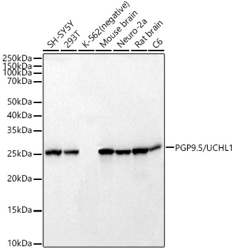
Western blot analysis of various lysates, using PGP9.5/UCHL1 Rabbit mAb (A20380) at 1:47000 dilution. Secondary antibody: HRP-conjugated Goat anti-Rabbit IgG (H+L) (AS014) at 1:10000 dilution. Lysates/proteins: 25µg per lane. Blocking buffer: 3% nonfat dry milk in TBST. Detection: ECL Basic Kit (RM00020). Exposure time: 1s. |
|
|
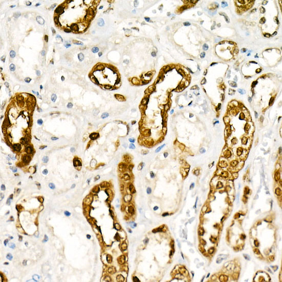
Immunohistochemistry analysis of paraffin-embedded Human kidney tissue using PGP9.5/UCHL1 Rabbit mAb (A20380) at a dilution of 1:800 (40x lens). High pressure antigen retrieval performed with 0.01M Citrate buffer (pH 6.0) prior to IHC staining. |
|
|
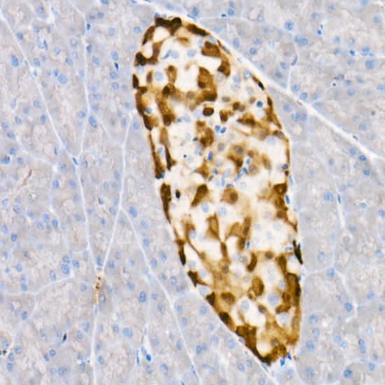
Immunohistochemistry analysis of paraffin-embedded Human pancreas tissue using PGP9.5/UCHL1 Rabbit mAb (A20380) at a dilution of 1:800 (40x lens). High pressure antigen retrieval performed with 0.01M Citrate buffer (pH 6.0) prior to IHC staining. |
|
|
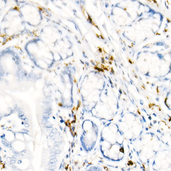
Immunohistochemistry analysis of paraffin-embedded Human tonsil tissue using PGP9.5/UCHL1 Rabbit mAb (A20380) at a dilution of 1:800 (40x lens). High pressure antigen retrieval performed with 0.01M Citrate buffer (pH 6.0) prior to IHC staining. |
|
|
Immunohistochemistry analysis of paraffin-embedded Mouse spleen tissue using PGP9.5/UCHL1 Rabbit mAb (A20380) at a dilution of 1:800 (40x lens). High pressure antigen retrieval performed with 0.01M Citrate buffer (pH 6.0) prior to IHC staining. |
|
|
Immunohistochemistry analysis of paraffin-embedded Rat pancreas tissue using PGP9.5/UCHL1 Rabbit mAb (A20380) at a dilution of 1:800 (40x lens). High pressure antigen retrieval performed with 0.01M Citrate buffer (pH 6.0) prior to IHC staining. |
|
|
Immunohistochemistry analysis of paraffin-embedded Rat spleen tissue using PGP9.5/UCHL1 Rabbit mAb (A20380) at a dilution of 1:800 (40x lens). High pressure antigen retrieval performed with 0.01M Citrate buffer (pH 6.0) prior to IHC staining. |
|
|
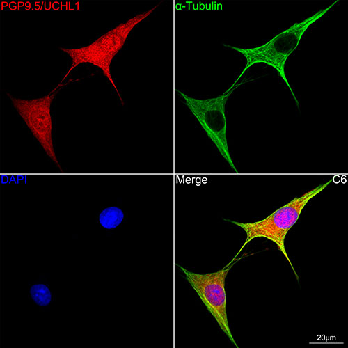
Confocal imaging of C6 cells using PGP9.5/UCHL1 Rabbit mAb (A20380,dilution 1:1000)(Red). The cells were counterstained with alpha-Tubulin Mouse mAb (AC012,dilution 1:400) (Green). DAPI was used for nuclear staining (blue). Objective: 60x. |
|
|
Immunofluorescence analysis of Mouse brain tissue using PGP9.5/UCHL1 Rabbit mAb (A20380) at a dilution of 1:1000 (40x lens). Secondary antibody: Cy3-conjugated Goat anti-Rabbit IgG (H+L)(AS007) at 1:500 dilution. Blue: DAPI for nuclear staining. Microwave antigen retrieval performed with 0.01M Citrate Buffer(pH 6.0) prior to IF staining. |
Produktgarantie und fachkundiger Support
