
[KD Validated] LAMP1/CD107a Rabbit mAb, Unconjugated, Monoclonal
Artikelnummer:
ABB-A21194
- Bilder (9)

| Artikelname: | [KD Validated] LAMP1/CD107a Rabbit mAb, Unconjugated, Monoclonal |
| Artikelnummer: | ABB-A21194 |
| Hersteller Artikelnummer: | A21194 |
| Alternativnummer: | ABB-A21194-100UL,ABB-A21194-20UL,ABB-A21194-500UL,ABB-A21194-1000UL |
| Hersteller: | ABclonal |
| Wirt: | Rabbit |
| Kategorie: | Antikörper |
| Applikation: | ELISA, FC, IF, IHC-P, WB |
| Spezies Reaktivität: | Human |
| Immunogen: | Recombinant protein (or fragment).This information is considered to be commercially sensitive. |
| Konjugation: | Unconjugated |
| Alternative Synonym: | LAMPA, CD107a, LGP120 |
| The protein encoded by this gene is a member of a family of membrane glycoproteins. This glycoprotein provides selectins with carbohydrate ligands. It may also play a role in tumor cell metastasis. |
| Klonalität: | Monoclonal |
| Klon-Bezeichnung: | [ARC52154] |
| Molekulargewicht: | 38kDa/45kDa |
| NCBI: | 3916 |
| UniProt: | P11279 |
| Reinheit: | Affinity purification |
| Sequenz: | AMFMVKNGNGTACIMANFSAAFSVNYDTKSGPKNMTFDLPSDATVVLNRSSCGKENTSDPSLVIAFGRGHTLTLNFTRNATRYSVQLMSFVYNLSDTHLFPNASSKEIKTVESITDIRADIDKKYRCVSGTQVHMNNVTVTLHDATIQAYLSNSSFSRGETRCEQDRPSPTTAPPAPPSPSPSPVPKSPSVDKYNVSGTNGTCLLASMGLQLNLTYERKDNTTVTRLLNINPNKTSASGSCGAHLVTLELHSEGT |
| Target-Kategorie: | LAMP1 |
| Antibody Type: | Primary Antibody |
| Application Verdünnung: | WB,1:10000 - 1:40000|IHC-P,1:500 - 1:5000|IF/ICC,1:200 - 1:2000|FC (intra),1:100 - 1:500|ELISA,Recommended starting concentration is 1 µg/mL. Please optimize the concentration based on your specific assay requirements. |
| Anwendungsbeschreibung: | Cross-Reactivity: Human. ResearchArea: Cancer,Signal Transduction,Cell Biology Developmental Biology,Autophagy,Endocrine Metabolism,Immunology Inflammation,CDs,Neuroscience, Cell Type Marker,Stem Cells,Hematopoietic Progenitors,Neuron marker. Shipping: Ice Bag |
|
|
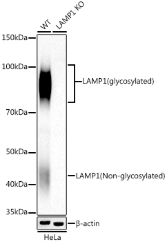
Western blot analysis of lysates from wild type (WT) and LAMP1 knockdown (KD) HeLa cells, using [KD Validated] LAMP1/CD107a Rabbit mAb (A21194) at 1:10000 dilution. Secondary antibody: HRP-conjugated Goat anti-Rabbit IgG (H+L) (AS014) at 1:10000 dilution. Lysates/proteins: 25µg per lane. Blocking buffer: 3% nonfat dry milk in TBST. Detection: ECL Basic Kit (RM00020). Exposure time: 20s. |
|
|
Immunohistochemistry analysis of paraffin-embedded Human colon carcinoma tissue using [KD Validated] LAMP1/CD107a Rabbit mAb (A21194) at a dilution of 1:2000 (40x lens). High pressure antigen retrieval performed with 0.01M Tris-EDTA Buffer(pH 9.0) prior to IHC staining. |
|
|
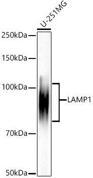
Western blot analysis of lysates from U-251MG cells, using [KD Validated] LAMP1/CD107a Rabbit mAb (A21194) at 1:10000 dilution. Secondary antibody: HRP-conjugated Goat anti-Rabbit IgG (H+L) (AS014) at 1:10000 dilution. Lysates/proteins: 25µg per lane. Blocking buffer: 3% nonfat dry milk in TBST. Detection: ECL Basic Kit (RM00020). Exposure time: 20s. |
|
|
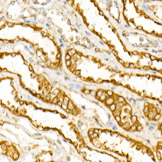
Immunohistochemistry analysis of paraffin-embedded Human kidney tissue using [KD Validated] LAMP1/CD107a Rabbit mAb (A21194) at a dilution of 1:2000 (40x lens). High pressure antigen retrieval performed with 0.01M Tris-EDTA Buffer(pH 9.0) prior to IHC staining. |
|
|
Immunohistochemistry analysis of paraffin-embedded Human tonsil tissue using [KD Validated] LAMP1/CD107a Rabbit mAb (A21194) at a dilution of 1:2000 (40x lens). High pressure antigen retrieval performed with 0.01M Tris-EDTA Buffer(pH 9.0) prior to IHC staining. |
|
|
Immunohistochemistry analysis of paraffin-embedded HeLa and HeLa-LAMP1-KO cells using [KD Validated] LAMP1/CD107a Rabbit mAb (A21194) at a dilution of 1:2200 (40x lens). High pressure antigen retrieval performed with 0.01M Tris-EDTA Buffer (pH 9.0) prior to IHC staining. |
|
|
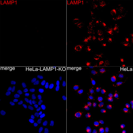
Confocal imaging of HeLa and HeLa-LAMP1-KD cells using [KD Validated] LAMP1/CD107a Rabbit mAb (A21194,dilution 1:200) followed by a further incubation with Cy3 Goat Anti-Rabbit IgG (H+L) (AS007,dilution 1:500)(Red).DAPI was used for nuclear staining (Blue). Objective: 100x. |
|
|
Flow cytometry: 1X10 6 knockdown (KD) HeLa cells (negative control,left)and HeLa cells (right) were intracellularly-stained with [KD Validated] LAMP1/CD107a Rabbit mAb (A21194,2.5 µg/mL,orange line) or ABflo 488 Rabbit IgG isotype control (A22069,5 µl/Test,blue line), followed by FITC conjugated goat anti-Rabbit pAb staining. Non-fluorescently stained cells were used as blank control (red line). |
|
|
Flow cytometry: 1X10 6 HeLa cells were intracellularly-stained with ABflo 488 Rabbit IgG isotype control (A22069,5 µl/Test,left) or [KD Validated] LAMP1/CD107a Rabbit mAb (A21194,2.5 µg/mL,right). |
Produktgarantie und fachkundiger Support
