
KAP1/TRIM28 Rabbit pAb, Unconjugated, Polyclonal
Artikelnummer:
ABB-A2245
- Bilder (9)
| Artikelname: | KAP1/TRIM28 Rabbit pAb, Unconjugated, Polyclonal |
| Artikelnummer: | ABB-A2245 |
| Hersteller Artikelnummer: | A2245 |
| Alternativnummer: | ABB-A2245-100UL,ABB-A2245-20UL |
| Hersteller: | ABclonal |
| Wirt: | Rabbit |
| Kategorie: | Antikörper |
| Applikation: | ELISA, IF, IHC-P, IP, WB |
| Spezies Reaktivität: | Human |
| Immunogen: | This information is considered to be commercially sensitive. |
| Konjugation: | Unconjugated |
| Alternative Synonym: | KAP1, TF1B, RNF96, TIF1B, PPP1R157, TIF1beta, KAP1/TRIM28 |
| The protein encoded by this gene mediates transcriptional control by interaction with the Kruppel-associated box repression domain found in many transcription factors. The protein localizes to the nucleus and is thought to associate with specific chromatin regions. The protein is a member of the tripartite motif family. This tripartite motif includes three zinc-binding domains, a RING, a B-box type 1 and a B-box type 2, and a coiled-coil region. |
| Application Verdünnung: | WB,1:1000 - 1:5000|IP,0.5µg-4µg antibody for 200µg-400µg extracts of whole cells|IF/ICC,1:50 - 1:200|IHC-P,1:50 - 1:200|ELISA,Recommended starting concentration is 1 µg/mL. Please optimize the concentration based on your specific assay requirements. |
| Anwendungsbeschreibung: | Cross-Reactivity: Human,Mouse,Rat. ResearchArea: Epigenetics Nuclear Signaling,Transcription Factors,Protein phosphorylation,Cancer,Invasion and Metastasis,Cell Biology Developmental Biology,Apoptosis,Ubiquitin. Shipping: Ice Bag |
|
|
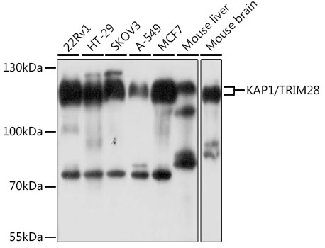
Western blot analysis of various lysates using KAP1/TRIM28 Rabbit pAb (A2245) at 1:1000 dilution. Secondary antibody: HRP-conjugated Goat anti-Rabbit IgG (H+L) (AS014) at 1:10000 dilution. Lysates/proteins: 25µg per lane. Blocking buffer: 3% nonfat dry milk in TBST. Detection: ECL Basic Kit (RM00020). Exposure time: 5s. |
|
|
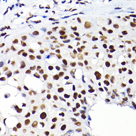
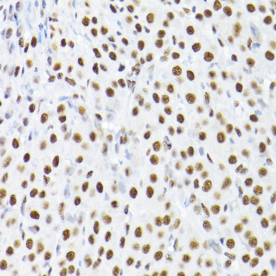
Immunohistochemistry analysis of paraffin-embedded Human breast cancer using KAP1/KAP1/TRIM28 Rabbit pAb (A2245) at dilution of 1:200 (40x lens). High pressure antigen retrieval performed with 0.01M Citrate buffer (pH 6.0) prior to IHC staining. |
|
|
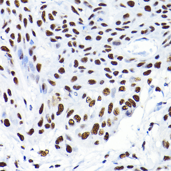
Immunohistochemistry analysis of paraffin-embedded Human esophageal cancer using KAP1/KAP1/TRIM28 Rabbit pAb (A2245) at dilution of 1:200 (40x lens). High pressure antigen retrieval performed with 0.01M Citrate buffer (pH 6.0) prior to IHC staining. |
|
|
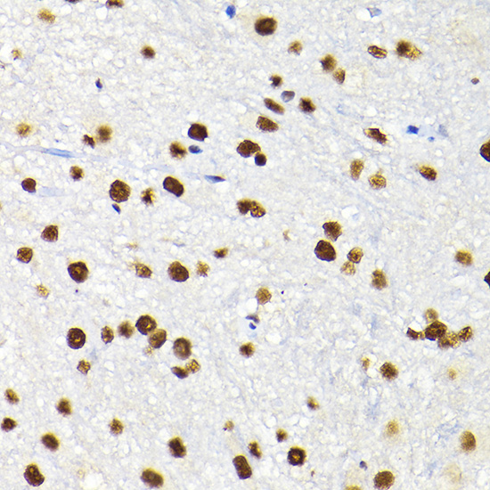
Immunohistochemistry analysis of paraffin-embedded Mouse spinal cord using KAP1/KAP1/TRIM28 Rabbit pAb (A2245) at dilution of 1:200 (40x lens). High pressure antigen retrieval performed with 0.01M Citrate buffer (pH 6.0) prior to IHC staining. |
|
|
Immunohistochemistry analysis of paraffin-embedded Rat ovary using KAP1/KAP1/TRIM28 Rabbit pAb (A2245) at dilution of 1:200 (40x lens). High pressure antigen retrieval performed with 0.01M Citrate buffer (pH 6.0) prior to IHC staining. |
|
|
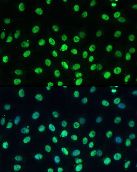
Immunofluorescence analysis of C6 cells using KAP1/TRIM28 Rabbit pAb (A2245) at dilution of 1:100 (40x lens). Secondary antibody: Cy3-conjugated Goat anti-Rabbit IgG (H+L) (AS007) at 1:500 dilution. Blue: DAPI for nuclear staining. |
|
|
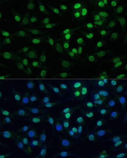
Immunofluorescence analysis of L929 cells using KAP1/TRIM28 Rabbit pAb (A2245) at dilution of 1:100 (40x lens). Secondary antibody: Cy3-conjugated Goat anti-Rabbit IgG (H+L) (AS007) at 1:500 dilution. Blue: DAPI for nuclear staining. |
|
|
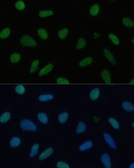
Immunofluorescence analysis of U-2 OS cells using KAP1/TRIM28 Rabbit pAb (A2245) at dilution of 1:100 (40x lens). Secondary antibody: Cy3-conjugated Goat anti-Rabbit IgG (H+L) (AS007) at 1:500 dilution. Blue: DAPI for nuclear staining. |
|
|
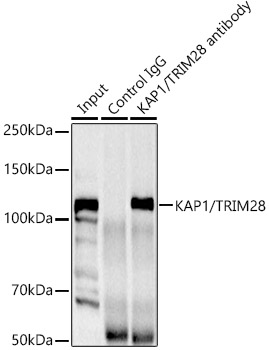
Immunoprecipitation analysis of 300ug extracts of HeLa cells using 3ug KAP1/TRIM28 Rabbit pAb (A2245 1:400). Western blot was performed from the immunoprecipitate using KAP1/TRIM28 Rabbit pAb (A2245) at a dilition of 1:1000. |
Produktgarantie und fachkundiger Support
