
Phospho-HDAC5-S498 Rabbit pAb, Unconjugated
Artikelnummer:
ABB-AP0202
- Bilder (1)
| Artikelname: | Phospho-HDAC5-S498 Rabbit pAb, Unconjugated |
| Artikelnummer: | ABB-AP0202 |
| Hersteller Artikelnummer: | AP0202 |
| Alternativnummer: | ABB-AP0202-100UL, ABB-AP0202-20UL |
| Hersteller: | ABclonal |
| Wirt: | Rabbit |
| Kategorie: | Antikörper |
| Applikation: | ELISA, WB |
| Spezies Reaktivität: | Human |
| Immunogen: | Synthetic peptide. This information is considered to be commercially sensitive. |
| Konjugation: | Unconjugated |
| Alternative Synonym: | HD5, NY-CO-9, Phospho-HDAC5-S498 |
| Histones play a critical role in transcriptional regulation, cell cycle progression, and developmental events. Histone acetylation/deacetylation alters chromosome structure and affects transcription factor access to DNA. The protein encoded by this gene belongs to the class II histone deacetylase/acuc/apha family. It possesses histone deacetylase activity and represses transcription when tethered to a promoter. It coimmunoprecipitates only with HDAC3 family member and might form multicomplex proteins. It also interacts with myocyte enhancer factor-2 (MEF2) proteins, resulting in repression of MEF2-dependent genes. This gene is thought to be associated with colon cancer. Two transcript variants encoding different isoforms have been found for this gene. |
| Application Verdünnung: | WB,1:500 - 1:2000|ELISA,Recommended starting concentration is 1 µg/mL. Please optimize the concentration based on your specific assay requirements. |
| Anwendungsbeschreibung: | Cross-Reactivity: Human,Mouse,Rat, ResearchArea: Epigenetics Nuclear Signaling,Nuclear Receptor Signaling,Protein phosphorylation,Signal Transduction,Cell Biology Developmental Biology,Cell Cycle,Cell Cycle Control-G1 S Checkpoint,Wnt -Catenin Signaling Pathway,Immunology Inflammation,NF-kB Signaling Pathway,Stem Cells,Cardiovascular,Heart,Hypertrophy. |
|
|
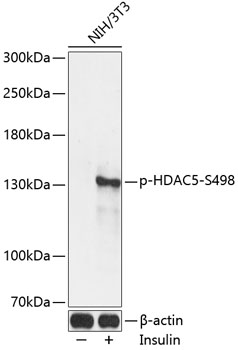
Western blot analysis of lysates from NIH/3T3 cells, using Phospho-HDAC5-S498 Rabbit pAb (AP0202) at 1:2000 dilution. NIH/3T3 cells were treated with Insulin (100nM) for 10 minutes after serum-starvation overnight. Secondary antibody: HRP-conjugated Goat anti-Rabbit IgG (H+L) (AS014) at 1:10000 dilution. Lysates/proteins: 25µg per lane. Blocking buffer: 3% BSA. Detection: ECL Enhanced Kit (RM00021). Exposure time: 30s. |
Produktgarantie und fachkundiger Support
