
Anti-SEMA3E, Rabbit, Polyclonal
Artikelnummer:
ATA-HPA029419
- Bilder (10)

| Artikelname: | Anti-SEMA3E, Rabbit, Polyclonal |
| Artikelnummer: | ATA-HPA029419 |
| Hersteller Artikelnummer: | HPA029419 |
| Alternativnummer: | ATA-HPA029419-100,ATA-HPA029419-25 |
| Hersteller: | Atlas Antibodies |
| Wirt: | Rabbit |
| Kategorie: | Antikörper |
| Applikation: | IHC |
| Spezies Reaktivität: | Human, Mouse |
| Immunogen: | Recombinant Protein Epitope Signature Tag (PrEST) antigen sequence |
| Konjugation: | Unconjugated |
| Alternative Synonym: | coll-5, KIAA0331, M-SemaK, SEMAH |
| sema domain, immunoglobulin domain (Ig), short basic domain, secreted, (semaphorin) 3E |
| Anti-SEMA3E |
| Klonalität: | Polyclonal |
| Konzentration: | 0.1 mg/ml |
| Isotyp: | IgG |
| NCBI: | 9723 |
| UniProt: | O15041 |
| Puffer: | 40% glycerol and PBS (pH 7.2). 0.02% sodium azide is added as preservative. |
| Reinheit: | Affinity purified using the PrEST antigen as affinity ligand |
| Sequenz: | MDLGLLFLRLHKSDAGTYFCQTVEHSFVHTVRKITLEVVEEEKVEDMFNKDDEEDRHHRMPCPAQSSISQGAKPWYKEFLQLIGYSNFQRVEEYCEKVWCTDRKRKKLKMSPSKWKYANPQEKKLRSKPEHYR |
| Lagerung: | Store at +4°C for short term storage. Long time storage is recommended at -20°C. |
| Antibody Type: | Monoclonal Antibody |
| Application Verdünnung: | IHC: 1:200 - 1:500 |
|
|
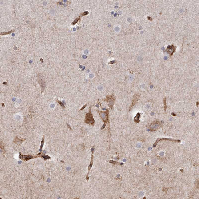
Immunohistochemical staining of human cerebral cortex shows weak cytoplasmic positivity in neurons. |
|
|
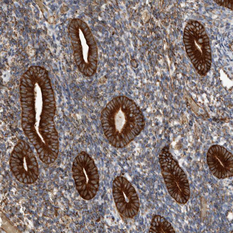
Immunohistochemical staining of human endometrium shows strong membranous positivity in glandular cells. |
|
|
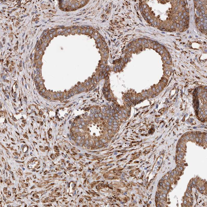
Immunohistochemical staining of mouse prostate shows moderate to strong membranous positivity in glandular cells. |
|
|
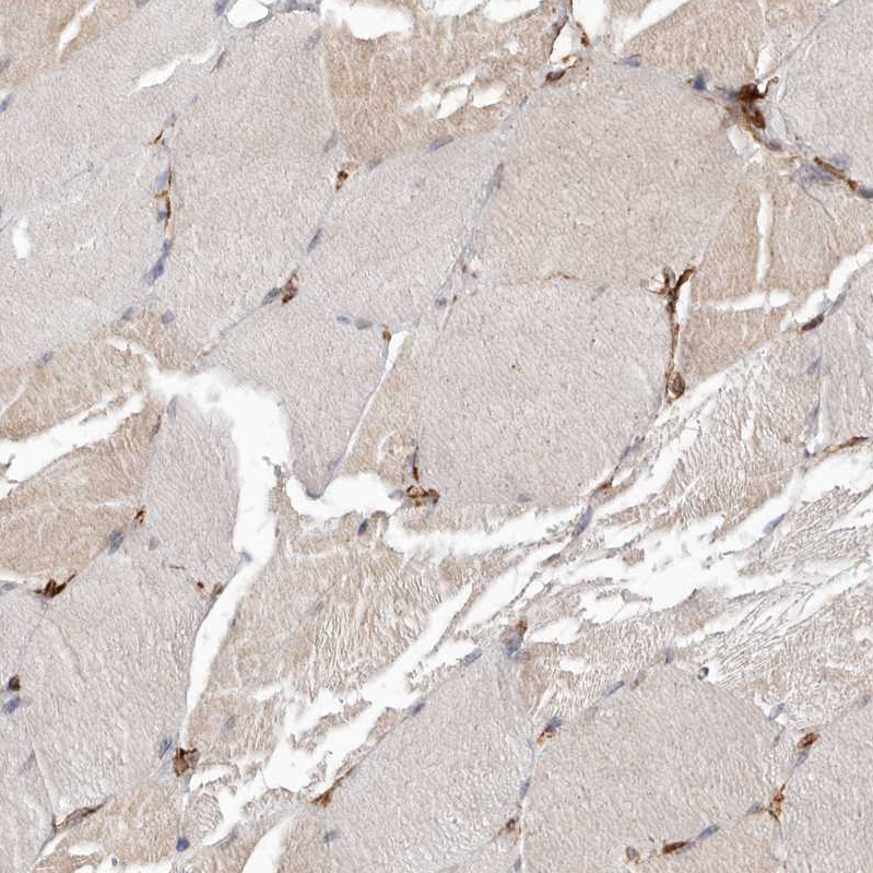
Immunohistochemical staining of human skeletal muscle shows no positivity in myocytes as expected. |
|
|
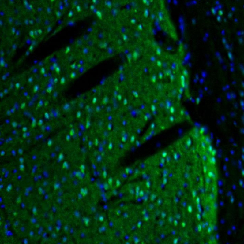
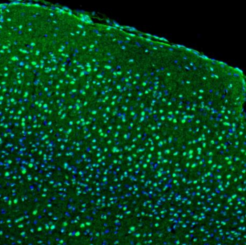
Immunofluorescence staining of mouse basal forebrain (caudate putamen) shows moderate to strong positivity |
|
|
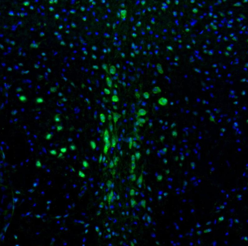
Immunofluorescence staining of human midbrain shows moderate to strong positivity of neurons in the dorsal raphe nucleus. |
|
|
Immunofluorescence staining of human cerebral cortex shows moderate to strong positivity of neurons in the visual cortex. |
|
|
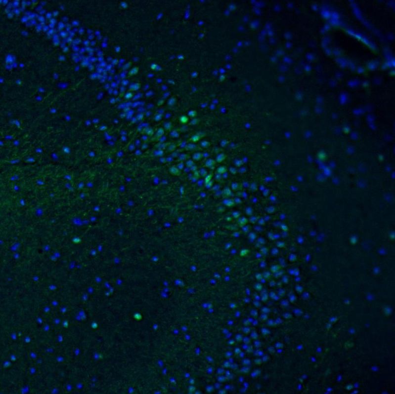
Immunofluorescence staining of mouse hippocampus shows moderate to strong positivity of neurons in the ca2. |
|
|
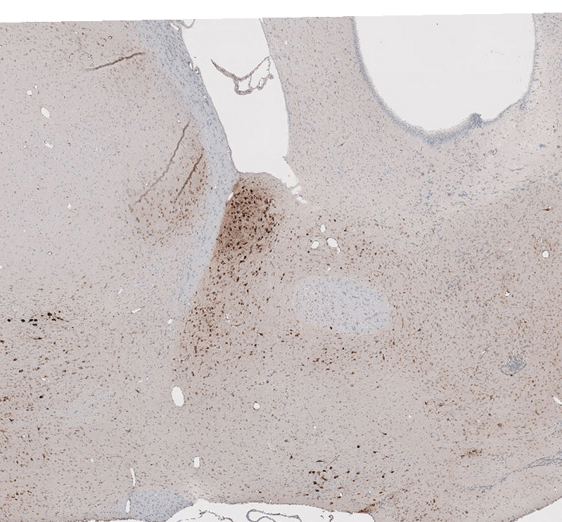
Immunohistochemical staining of mouse hypothalamus shows moderate to strong cytoplasmic positivity in neurons. |
|
|
Produktgarantie und fachkundiger Support
