Application Notes: For reconstitution, we recommend adding 100uL distilled water to a final antibody concentration of about 1 mg/mL. To use this carrier-free antibody for conjugation experiment, we strongly recommend performing another round of desalting process
HEK293T cells were transfected with the pCMV6-ENTRY control (Left lane) or pCMV6-ENTRY TP53 (RC200003, Right lane) cDNA for 48 hrs and lysed.
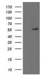
Western blot analysis of extracts (35ug) from 9 different cell lines by using anti-TP53 monoclonal antibody.
Immunohistochemical staining of paraffin-embedded Carcinoma of Human pancreas tissue using anti-TP53 mouse monoclonal antibody. (Heat-induced epitope retrieval by 10mM citric buffer, pH6.0, 100C for 10min)
Immunohistochemical staining of paraffin-embedded Carcinoma of Human bladder tissue using anti-TP53 mouse monoclonal antibody. (Heat-induced epitope retrieval by 10mM citric buffer, pH6.0, 100C for 10min)
Anti-TP53 mouse monoclonal antibody (TA502780) immunofluorescent staining of COS7 cells transiently transfected by pCMV6-ENTRY TP53)
HEK293T cells transfected with either RC200003 overexpress plasmid (Red) or empty vector control plasmid (Blue) were immunostained by anti-TP53 antibody (TA502780), and then analyzed by flow cytometry.
Flow cytometric Analysis of Hela cells, using anti-TP53 antibody (TA502780), (Red), compared to a nonspecific negative control antibody, (Blue).
Flow cytometric Analysis of Jurkat cells, using anti-TP53 antibody (TA502780), (Red), compared to a nonspecific negative control antibody, (Blue).
* Mehrwertsteuer und Versandkosten nicht enthalten. Irrtümer und Preisänderungen vorbehalten