VGLUT1 Antibody (Biotin), IgG1, Clone: [N28/9 Formerly sold as S28-9], Mouse, Monoclonal
Artikelnummer:
BYT-ORB149725
Hersteller Artikelnummer:
orb149725
Alternativnummer:
BYT-ORB149725-100
Hersteller:
Biorbyt
Wirt:
Mouse
Kategorie:
Antikörper
Applikation:
IHC, WB
Spezies Reaktivität:
Human, Mouse, Rat
Immunogen:
Fusion protein amino acids 493-560 (cytoplasmic C-terminus) of rat VGlut1
Konjugation:
Biotin
Alternative Synonym:
VGLUT1, Vesicular Glutamate Transporter 1, SLC17A7, BNPI, Solute Carrier Family 17 Member 7, Brain-specific Na(+)-dependent Inorganic Phosphate Cotransporter, Solute Carrier Family 17 (Sodium-Dependent Inorganic Phosphate Cotransporter), Member 7, Solute Carrier Family 17 (Vesicular Glutamate Transporter), Member 7, VGluT1, VGLUT 1
Mouse monoclonal to VGlut1 (Biotin). VGLUT1 is expressed in a subset of glutamate neurons and transports glutamate into native synaptic vesicles from the brain, exhibiting a conductance for chloride that is blocked by glutamate. Vesicular glutamate transport has a substantially lower apparent affinity than the plasma membrane excitatory amino acid transporters. Glutamate transport by VGLUT1 is saturated with a K(m) of approximately 2 mM, in the same range as transport by synaptic vesicles. Finally, plasma membrane glutamate transporters recognize both aspartate and glutamate as substrates, whereas VGLUT1 does not recognize aspartate..
136.36mM Ethanolamine, 133.23 mM Chlorides, 9.55mM Phosphates, 9.55mM Sodium Bicarbonate
Target-Kategorie:
VGLUT1
Application Verdünnung:
WB (1:1000)
Anwendungsbeschreibung:
Application Notes: 1 µg/ml of SMC-394 was sufficient for detection of VGLut1 in 20 µg of rat brain lysate by colorimetric immunoblot analysis using goat anti-mouse IgG:HRP as the secondary antibody
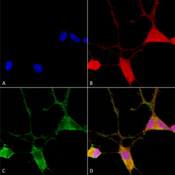
Immunocytochemistry/Immunofluorescence analysis using Mouse Anti-VGLUT1 Monoclonal Antibody, Clone N28/9. Tissue: Neuroblastoma cells (SH-SY5Y). Species: Human. Fixation: 4% PFA for 15 min. Primary Antibody: Mouse Anti-VGLUT1 Monoclonal Antibody at 1:100 for overnight at 4C with slow rocking. Secondary Antibody: AlexaFluor 488 at 1:1000 for 1 hour at RT. Counterstain: Phalloidin-iFluor 647 (red) F-Actin stain, Hoechst (blue) nuclear stain at 1:800, 1.6mM for 20 min at RT. (A) Hoechst (blue) nuclear stain. (B) Phalloidin-iFluor 647 (red) F-Actin stain. (C) VGLUT1 Antibody (D) Composite.
Immunohistochemistry analysis using Mouse Anti-VGLUT1 Monoclonal Antibody, Clone N28/9. Tissue: spinal cord. Species: Mouse. Fixation: 4% PFA. Primary Antibody: Mouse Anti-VGLUT1 Monoclonal Antibody at 1:500 for 16 hours at RT. Secondary Antibody: Alexa Fluor 555 Donkey Anti-Mouse (red) at 1:2000 for 2 hours at RT. Counterstain: NeuN neuronal stain (green). Magnification: 20X.
Western Blot analysis of Rat brain membrane lysate showing detection of VGLUT1 protein using Mouse Anti-VGLUT1 Monoclonal Antibody, Clone N28/9. Primary Antibody: Mouse Anti-VGLUT1 Monoclonal Antibody at 1:1000.
* Mehrwertsteuer und Versandkosten nicht enthalten. Irrtümer und Preisänderungen vorbehalten