136.36mM Ethanolamine, 133.23 mM Chlorides, 9.55mM Phosphates, 9.55mM Sodium Bicarbonate
Target-Kategorie:
GABA A Receptor
Application Verdünnung:
WB (1:1000), IHC (1:1000), ICC/IF (1:100)
Anwendungsbeschreibung:
Application Notes: 1 µg/ml of SMC-346 was sufficient for detection of alpha1 GABA receptor in 10 µg of rat brain lysate by colorimetric immunoblot analysis using Goat anti-mouse IgG:HRP as the secondary antibody
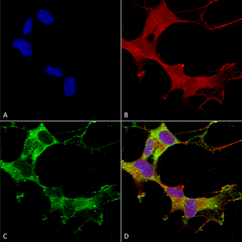
Immunocytochemistry/Immunofluorescence analysis using Mouse Anti-GABA-A Receptor Alpha-1 Monoclonal Antibody, Clone N95/35. Tissue: Neuroblastoma cells (SH-SY5Y). Species: Human. Fixation: 4% PFA for 15 min. Primary Antibody: Mouse Anti-GABA-A Receptor Alpha-1 Monoclonal Antibody at 1:50 for overnight at 4C with slow rocking. Secondary Antibody: AlexaFluor 488 at 1:1000 for 1 hour at RT. Counterstain: Phalloidin-iFluor 647 (red) F-Actin stain, Hoechst (blue) nuclear stain at 1:800, 1.6mM for 20 min at RT. (A) Hoechst (blue) nuclear stain. (B) Phalloidin-iFluor 647 (red) F-Actin stain. (C) GABA-A Receptor Alpha-1 Antibody (D) Composite.
Immunohistochemistry analysis using Mouse Anti-GABA A Receptor Monoclonal Antibody, Clone N95/35. Tissue: backskin. Species: Mouse. Fixation: Bouins Fixative and paraffin-embedded. Primary Antibody: Mouse Anti-GABA A Receptor Monoclonal Antibody at 1:100 for 1 hour at RT. Secondary Antibody: FITC Goat Anti-Mouse (green) at 1:50 for 1 hour at RT. Localization: Dermal Cells.
Western Blot analysis of Human Cell line lysates showing detection of GABA A Receptor protein using Mouse Anti-GABA A Receptor Monoclonal Antibody, Clone N95/35. Load: 15 µg. Block: 1.5% BSA for 30 minutes at RT. Primary Antibody: Mouse Anti-GABA A Receptor Monoclonal Antibody at 1:1000 for 2 hours at RT. Secondary Antibody: Sheep Anti-Mouse IgG: HRP for 1 hour at RT.
* Mehrwertsteuer und Versandkosten nicht enthalten. Irrtümer und Preisänderungen vorbehalten