Sample Tissue: Human 786-0 Whole Cell, Antibody dilution: 3 ug/ml.
Sample Tissue: Human HT1080 Whole Cell, Antibody dilution: 1 ug/ml.
Sample Tissue: Human Lung Tumor, Antibody dilution: 1 ug/ml.
Positive control (+): 293T (2T), Negative control (-): Lung tumor (T-LU), Antibody concentration: 5 ug/ml.
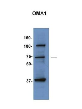
Lanes: Lane 1: 10 ug human fibroblast mitochondria, Lane 2: 15 ug fish embryo lysate, 6 h post fertilization, Lane 3: 30 ug fish embryo lysate, 6 days, Primary Antibody dilution: 1:1000, Secondary Antibody: Anti-Rabbit HRP, Secondary Antibody dilution: 1:5000, Gene Name: OMA1.
Surface Plasmon Resonance Kinetic Characterization of Polyclonal Antibody Affinity. Purified polyclonal antibodies were immobilized on a Protein A/G coated Carterra LSA sensor chip (PAGH200M) at concentrations of 5, and 50 ug/ml in duplicate. Antibodies on the surface were exposed to interaction with peptides sequentially via microfluidic controlled flow at 333nM peptide concentration for kinetic characterization of the binders for affinity and specificity, followed by curve fitting using the Kinetics software. Kd determinations for both concentrations were averaged and results and standard deviation are shown.