
Histone H3 Monoclonal Antibody, IgG2b, Clone: [1G2E4], Unconjugated, Mouse
Artikelnummer:
CSB-MA010418A0M
- Bilder (7)
| Artikelname: | Histone H3 Monoclonal Antibody, IgG2b, Clone: [1G2E4], Unconjugated, Mouse |
| Artikelnummer: | CSB-MA010418A0M |
| Hersteller Artikelnummer: | CSB-MA010418A0m |
| Alternativnummer: | CSB-MA010418A0M-100UL, CSB-MA010418A0M-50UL |
| Hersteller: | Cusabio |
| Wirt: | Mouse |
| Kategorie: | Antikörper |
| Applikation: | ELISA, IHC, IP, WB |
| Spezies Reaktivität: | Human, Mouse, Rabbit, Rat |
| Konjugation: | Unconjugated |
| Alternative Synonym: | Histone H3 |
| Klonalität: | Monoclonal |
| Klon-Bezeichnung: | [1G2E4] |
| Isotyp: | IgG2b |
| Puffer: | Preservative: 0.03% Proclin 300<br />Constituents: 50% Glycerol, 0.01M PBS, PH 7.4 |
| Reinheit: | >95%, Protein A purified |
| Formulierung: | Liquid |
| Target-Kategorie: | Histone H3 |
| Application Verdünnung: | Recommended dilution: WB:1:5000-1:640000, IHC: 1:100-1:200, IP:2µl-5µl |
|
|
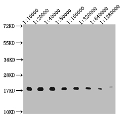
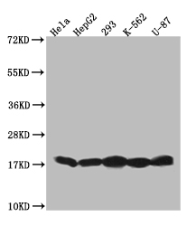
Western Blot Positive WB detected in: 10ug Hela whole cell lysate, HistoneH3 antibody at 1:10000, 1:20000, 1:40000, 1:80000, 1:160000, 1:320000, 1:640000, 1:1280000 Secondary Goat polyclonal to mouse IgG at 1/50000 dilution Predicted band size: 15-25 KDa Observed band size: 15-25 KDa Exposure time:5s |
|
|
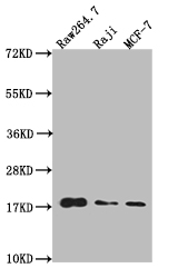
Western Blot Positive WB detected in: Raw264.7 whole cell lysate, Raji whole cell lysate, MCF-7 whole cell lysate All lanes: HistoneH3 antibody at 1:20000 Secondary Goat polyclonal to mouse IgG at 1/50000 dilution Predicted band size: 15-25 KDa Observed band size: 15-25 KDa Exposure time: 1min |
|
|
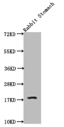
Western Blot Positive WB detected in: Rabbit stomach tissue All lanes: HistoneH3 antibody at 1:20000 Secondary Goat polyclonal to mouse IgG at 1/50000 dilution Predicted band size: 15-25 KDa Observed band size: 15-25 KDa Exposure time: 1min |
|
|
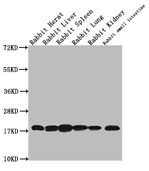
Western Blot Positive WB detected in: Rabbit heart tissue, Rabbit liver tissue,Rabbit spleen tissue, Rabbit lung tissue, Rabbit kidney tissue, Rabbit small intestine tissue All lanes: HistoneH3 antibody at 1:20000 Secondary Goat polyclonal to mouse IgG at 1/50000 dilution Predicted band size: 15-25 KDa Observed band size: 15-25 KDa Exposure time: 2min |
|
|
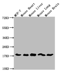
Western Blot Positive WB detected in: MCF-7 whole cell lysate, Mouse heart tissue, Mouse liver tissue, Mouse lung tissue, Mouse brain tissue tissue All lanes: HistoneH3 antibody at 1:5000 Secondary Goat polyclonal to mouse IgG at 1/50000 dilution Predicted band size: 15-25 KDa Observed band size: 15-25 KDa Exposure time: 5s |
|
|
CSB-MA010418A0m |
|
|
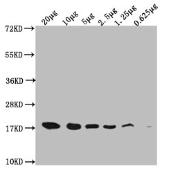
Western Blot Positive WB detected in: Hela whole cell lysate at 10ug, 5ug, 2.5ug, 1.25ug, 0.625ug All lanes: HistoneH3 antibody at 1:5000 Secondary Goat polyclonal to mouse IgG at 1/50000 dilution Predicted band size: 15-25 KDa Observed band size: 15-25 KDa Exposure time: 5s |
Produktgarantie und fachkundiger Support
