
KCNQ3 Antibody, Unconjugated, Rabbit, Polyclonal
Artikelnummer:
CSB-PA012091LA01HU
- Bilder (6)

| Artikelname: | KCNQ3 Antibody, Unconjugated, Rabbit, Polyclonal |
| Artikelnummer: | CSB-PA012091LA01HU |
| Hersteller Artikelnummer: | CSB-PA012091LA01HU |
| Alternativnummer: | CSB-PA012091LA01HU-100UG, CSB-PA012091LA01HU-50UG |
| Hersteller: | Cusabio |
| Wirt: | Rabbit |
| Kategorie: | Antikörper |
| Applikation: | ELISA, IF, WB |
| Spezies Reaktivität: | Human, Mouse, Rat |
| Konjugation: | Unconjugated |
| Alternative Synonym: | BFNC 2 antibody, BFNC antibody, BFNC2 antibody, EBN 2 antibody, EBN2 antibody, KCNQ 3 antibody, KCNQ3 antibody, KCNQ3_HUMAN antibody, KQT like 3 antibody, KQT-like 3 antibody, KV7.3 antibody, Potassium channel subunit alpha KvLQT3 antibody, Potassium channel voltage gated subfamily Q member 3 antibody, Potassium voltage gated channel KQT like protein 3 antibody, Potassium voltage gated channel KQT like subfamily member 3 antibody, Potassium voltage gated channel subfamily KQT member 3 antibody, Potassium voltage-gated channel subfamily KQT member 3 antibody, Voltage gated potassium channel subunit Kv7.3 antibody, Voltage-gated potassium channel subunit Kv7.3 antibody |
| Klonalität: | Polyclonal |
| UniProt: | O43525 |
| Puffer: | Preservative: 0.03% Proclin 300<br />Constituents: 50% Glycerol, 0.01M PBS, pH 7.4 |
| Reinheit: | Antigen Affinity Purified |
| Formulierung: | Liquid |
| Target-Kategorie: | KCNQ3 |
| Application Verdünnung: | Recommended dilution: WB:1:500-1:2000,IF:1:10-1:50 |
|
|
|
|
|
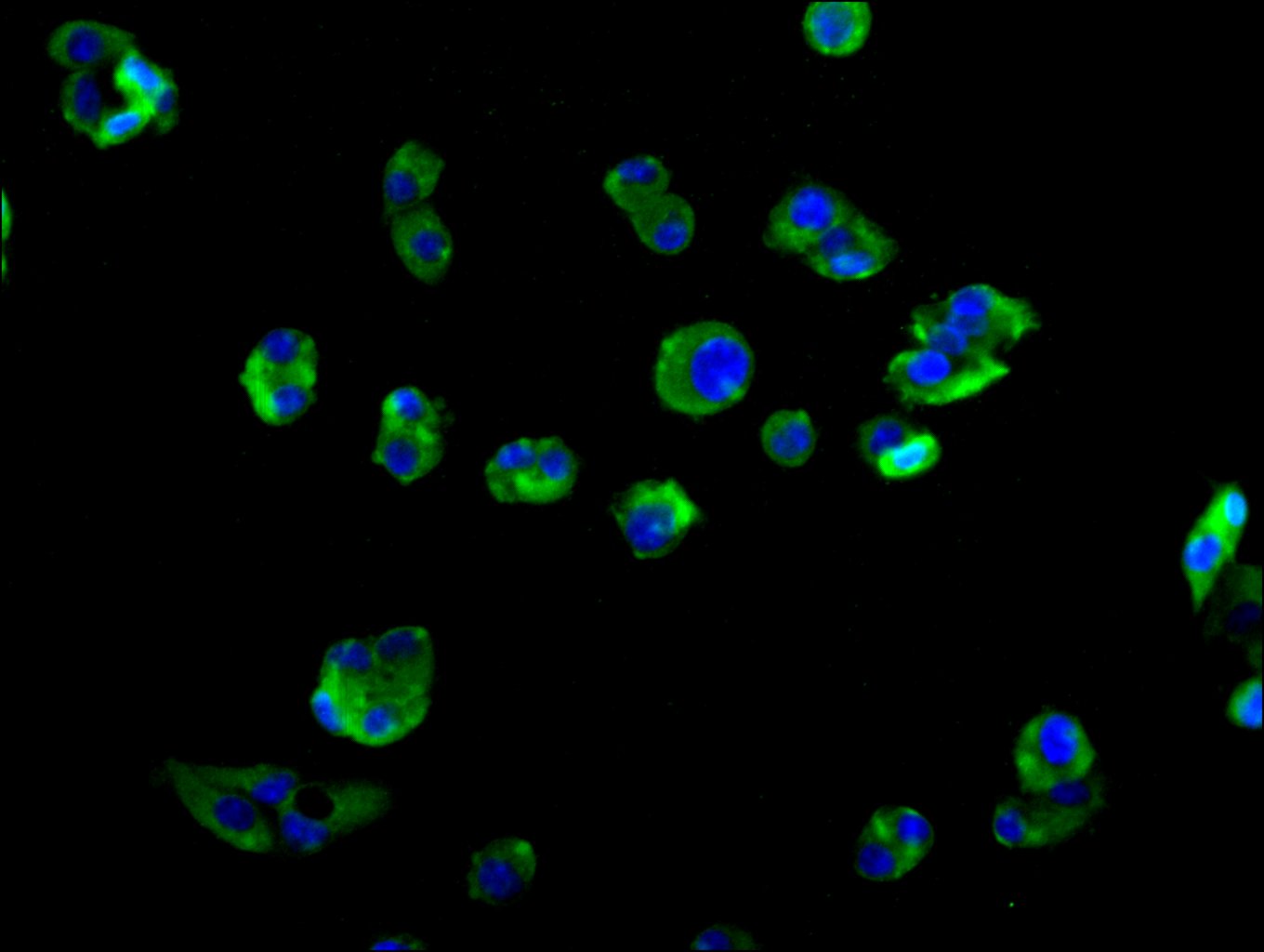
Immunofluorescence staining of HepG2 cell with CSB-PA012091LA01HU at 1:10, counter-stained with DAPI. The cells were fixed in 4% formaldehyde and blocked in 10% normal Goat Serum. The cells were then incubated with the antibody overnight at 4C. The secondary antibody was Alexa Fluor 488-congugated AffiniPure Goat Anti-Rabbit IgG(H+L). |
|
|
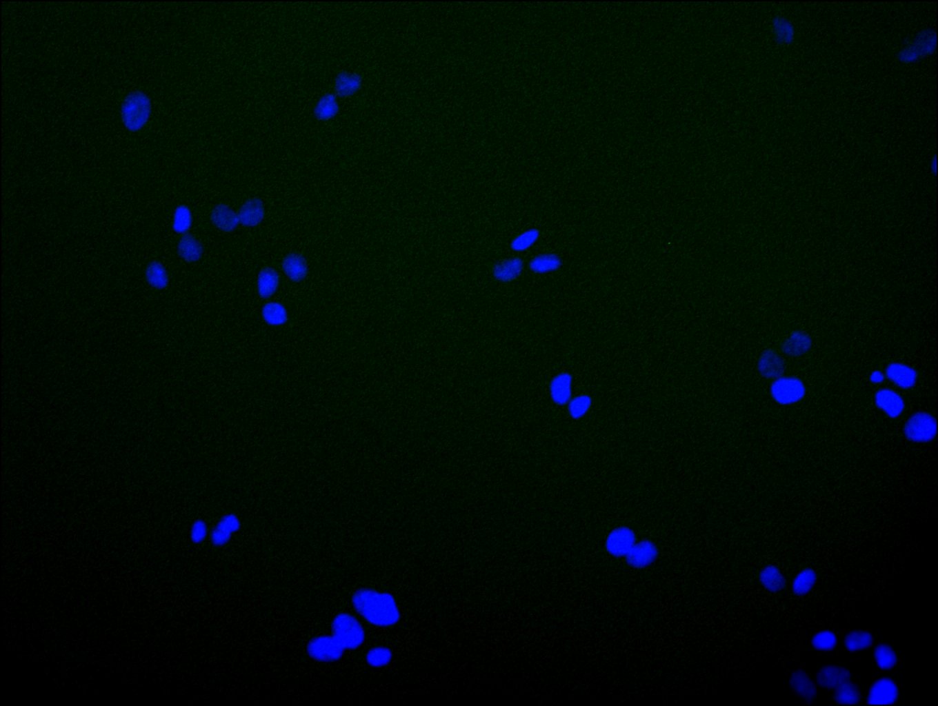
Immunofluorescence staining of HepG2 cell with 5% goat serum, counter-stained with DAPI. The cells were fixed in 4% formaldehyde and blocked in 10% normal Goat Serum. The cells were then incubated with the antibody overnight at 4C. The secondary antibody was Alexa Fluor 488-congugated AffiniPure Goat Anti-Rabbit IgG(H+L). |
|
|
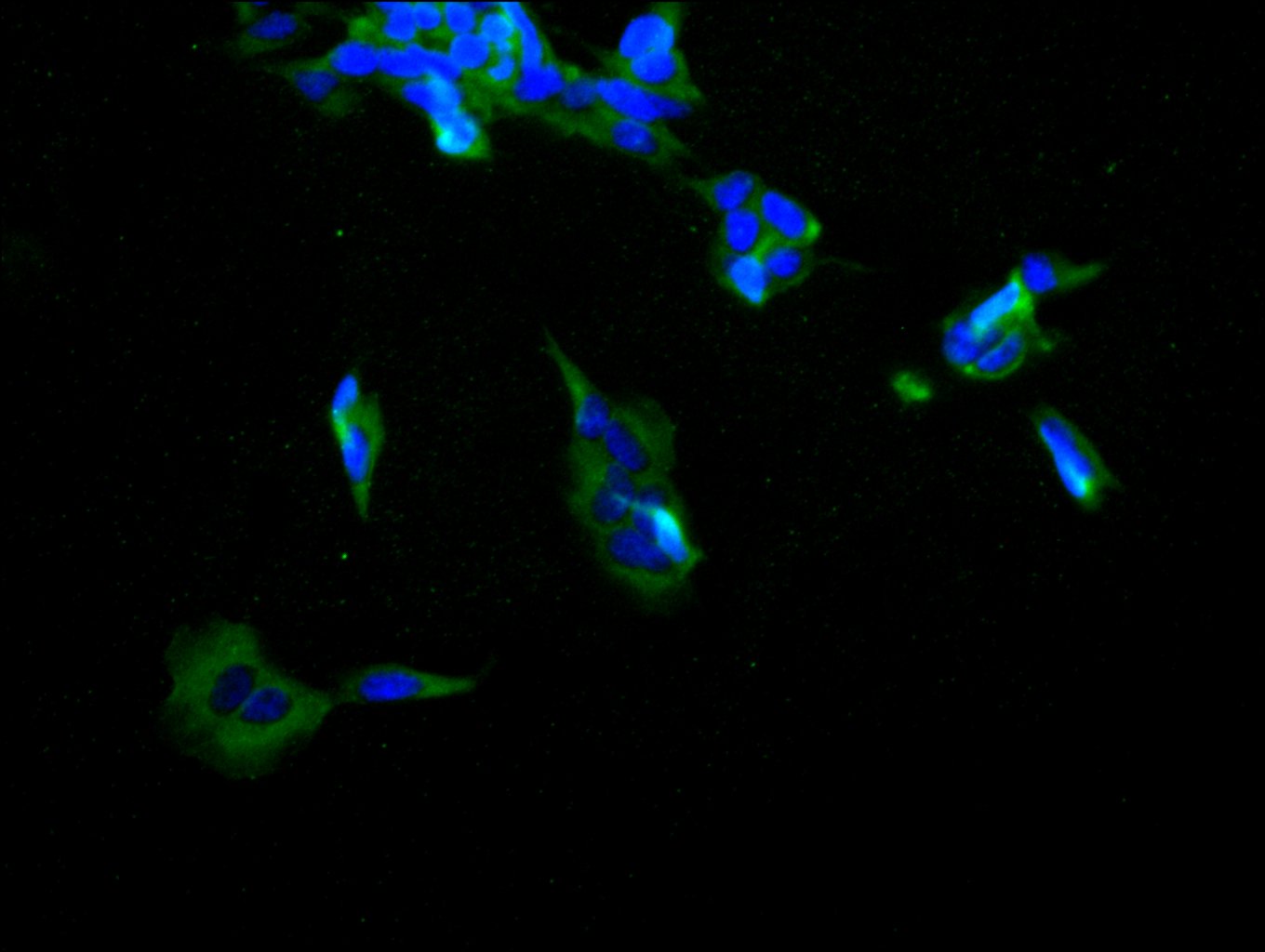
Immunofluorescence staining of SH-SY5Y cell with CSB-PA012091LA01HU at 1:10, counter-stained with DAPI. The cells were fixed in 4% formaldehyde and blocked in 10% normal Goat Serum. The cells were then incubated with the antibody overnight at 4C. The secondary antibody was Alexa Fluor 488-congugated AffiniPure Goat Anti-Rabbit IgG(H+L). |
|
|
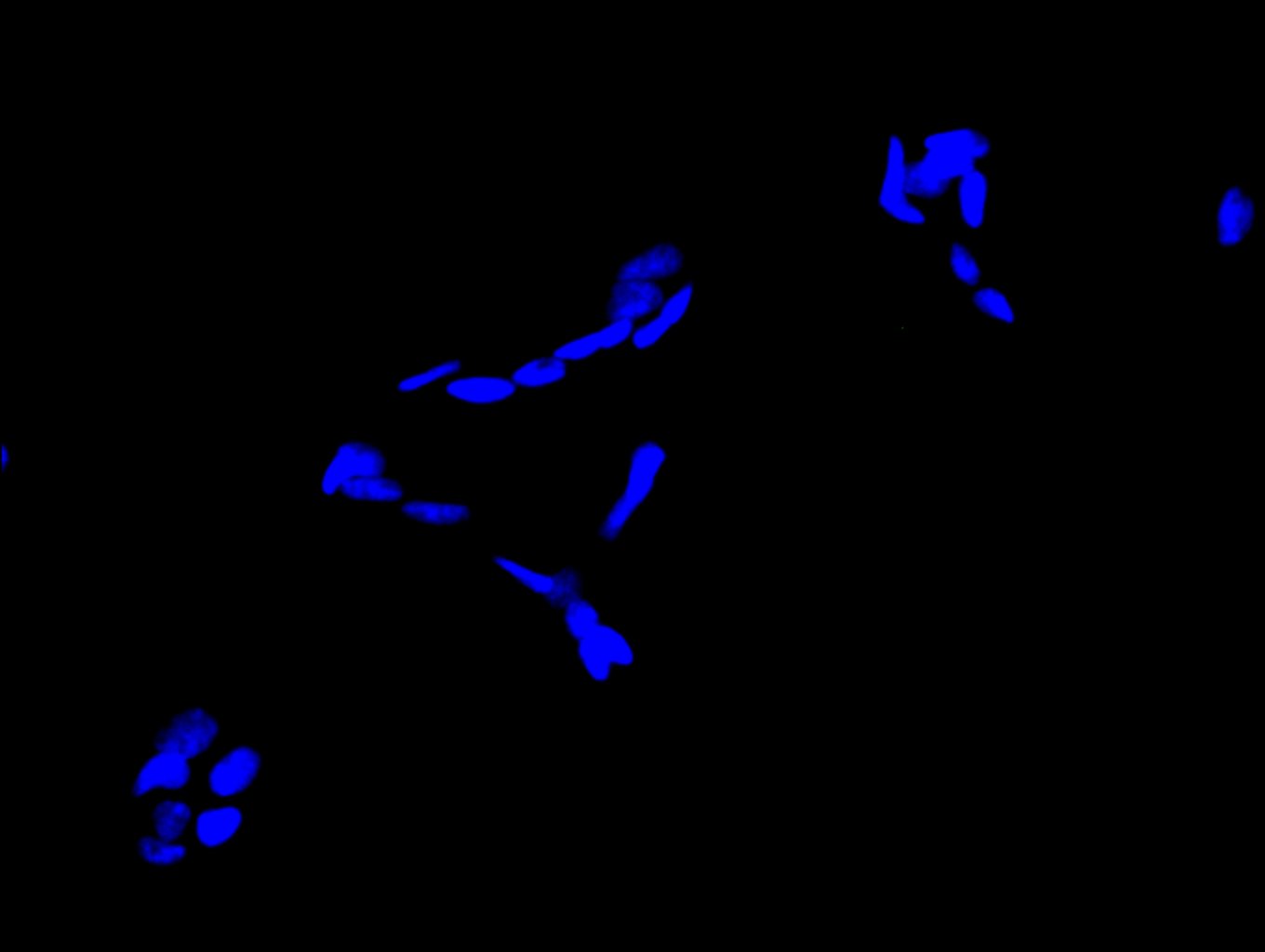
Immunofluorescence staining of SH-SY5Y cell with 5% goat serum, counter-stained with DAPI. The cells were fixed in 4% formaldehyde and blocked in 10% normal Goat Serum. The cells were then incubated with the antibody overnight at 4C. The secondary antibody was Alexa Fluor 488-congugated AffiniPure Goat Anti-Rabbit IgG(H+L). |
|
|
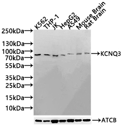
Western Blot Positive WB detected in: K562 whole cell lysate(30µg), THP-1 whole cell lysate(30µg), JK whole cell lysate(30µg), HepG2 whole cell lysate(30µg), A549 whole cell lysate(30µg), Mouse Brain tissue lysate(30µg),Rat Brain tissue lysate(30µg) All lanes: KCNQ3 antibody at 1:1000 Secondary Goat polyclonal to rabbit IgG at 1/20000 dilution Predicted band size: 97,85,90,100 kDa Observed band size: 100 kDa Exposure time:120s |
Produktgarantie und fachkundiger Support
