
SPOCK1 Antibody, Unconjugated, Rabbit, Polyclonal
Artikelnummer:
CSB-PA730785LA01HU
- Bilder (6)

| Artikelname: | SPOCK1 Antibody, Unconjugated, Rabbit, Polyclonal |
| Artikelnummer: | CSB-PA730785LA01HU |
| Hersteller Artikelnummer: | CSB-PA730785LA01HU |
| Alternativnummer: | CSB-PA730785LA01HU-100UG, CSB-PA730785LA01HU-50UG |
| Hersteller: | Cusabio |
| Wirt: | Rabbit |
| Kategorie: | Antikörper |
| Applikation: | ELISA, IHC, WB |
| Spezies Reaktivität: | Human, Mouse, Rat |
| Konjugation: | Unconjugated |
| Alternative Synonym: | SPOCK1 antibody, SPOCK antibody, TIC1 antibody, TICN1 antibody, Testican-1 antibody, Protein SPOCK antibody |
| Klonalität: | Polyclonal |
| UniProt: | Q08629 |
| Puffer: | Preservative: Liquid in PBS containing 50% glycerol, and 0.02% sodium azide. |
| Reinheit: | Antigen Affinity Purified |
| Formulierung: | Liquid |
| Target-Kategorie: | SPOCK1 |
| Application Verdünnung: | Recommended dilution: WB:1:500-1:2000, IHC:1:20-1:200 |
|
|
|
|
|
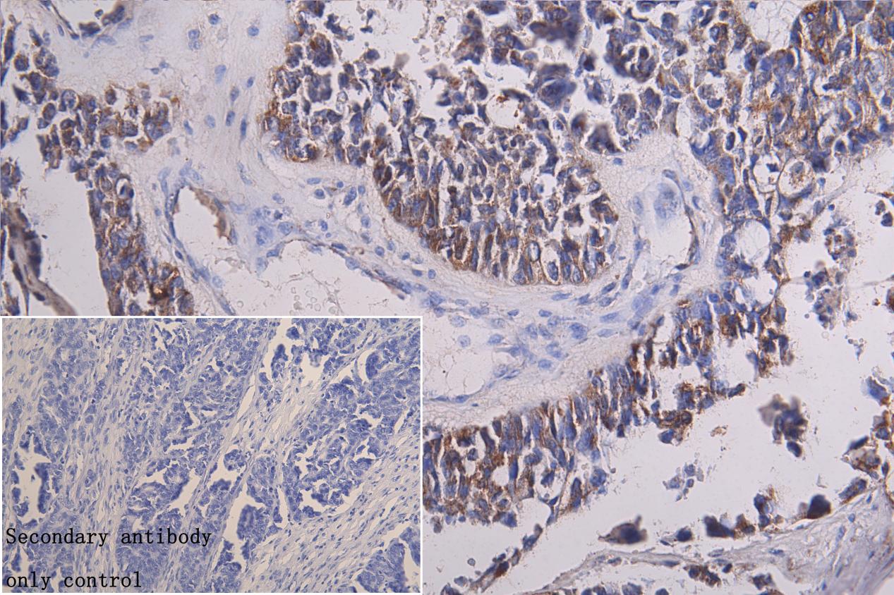
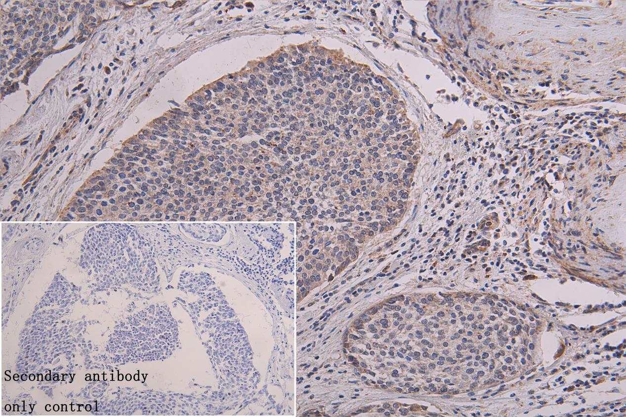
IHC image of CSB-PA730785LA01HU diluted at 1:100 and staining in paraffin-embedded human endometriall cancer tissue performed on a Leica BondTM system. After dewaxing and hydration, antigen retrieval was mediated by high pressure in a citrate buffer (pH 6.0). Section was blocked with 10% normal goat serum 30min at RT. Then primary antibody (1% BSA) was incubated at 4C overnight. The primary is detected by a Goat anti-rabbit polymer IgG labeled by HRP and visualized using 0.05% DAB. Secondary antibody only control: uses 1% BSA instead of primary antibody |
|
|
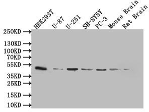
Western Blot Positive WB detected in: 293T whole cell lysate (20µg), U87 whole cell lysate (20µg), U251 whole cell lysate (20µg), SY5Y whole cell lysate (20µg), PC-3 whole cell lysate (20µg), Mouse brain tissue lysate (20µg), Rat brain tissue lysate (20µg) All lanes: SPOCK1 antibody at 1:1000 Secondary Goat polyclonal to rabbit IgG at 1/50000 dilution Predicted band size: 50 kDa Observed band size: 50 kDa |
|
|
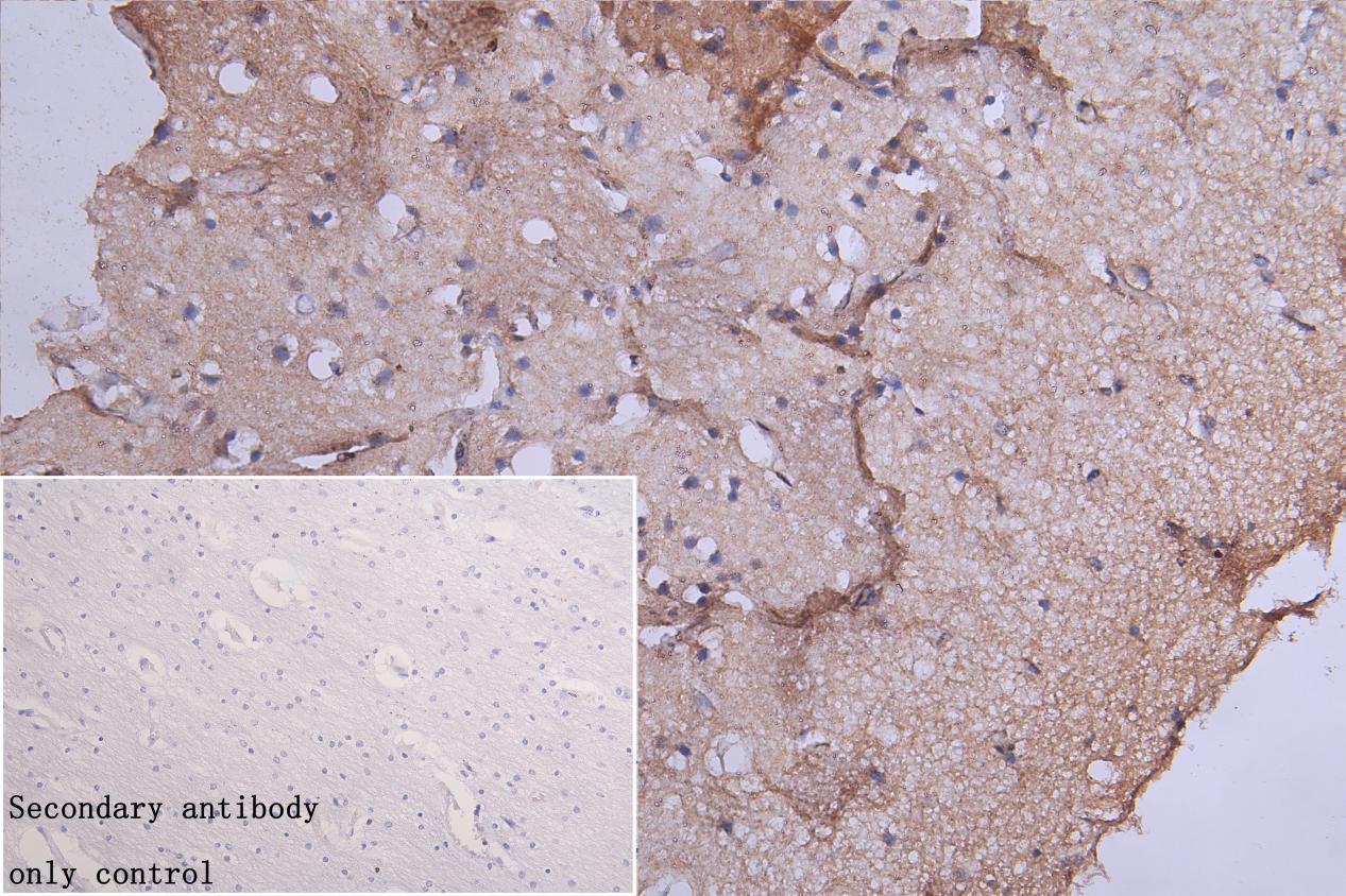
IHC image of CSB-PA730785LA01HU diluted at 1:100 and staining in paraffin-embedded human brain tissue performed on a Leica BondTM system. After dewaxing and hydration, antigen retrieval was mediated by high pressure in a citrate buffer (pH 6.0). Section was blocked with 10% normal goat serum 30min at RT. Then primary antibody (1% BSA) was incubated at 4C overnight. The primary is detected by a Goat anti-rabbit polymer IgG labeled by HRP and visualized using 0.05% DAB. Secondary antibody only control: uses 1% BSA instead of primary antibody |
|
|
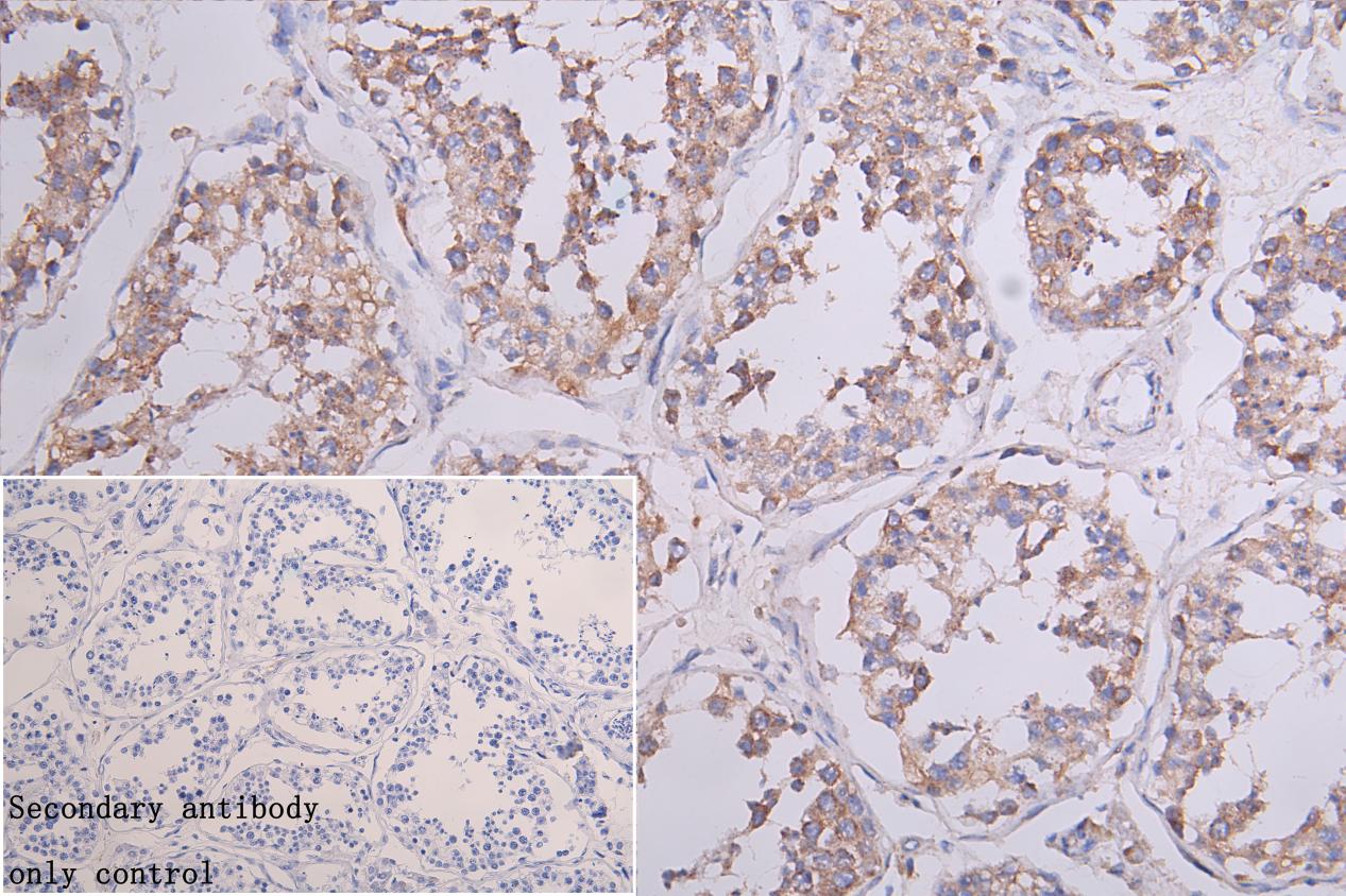
IHC image of CSB-PA730785LA01HU diluted at 1:100 and staining in paraffin-embedded human testis tissue performed on a Leica BondTM system. After dewaxing and hydration, antigen retrieval was mediated by high pressure in a citrate buffer (pH 6.0). Section was blocked with 10% normal goat serum 30min at RT. Then primary antibody (1% BSA) was incubated at 4C overnight. The primary is detected by a Goat anti-rabbit polymer IgG labeled by HRP and visualized using 0.05% DAB. Secondary antibody only control: uses 1% BSA instead of primary antibody |
|
|
IHC image of CSB-PA730785LA01HU diluted at 1:100 and staining in paraffin-embedded human Cervical cancer tissue performed on a Leica BondTM system. After dewaxing and hydration, antigen retrieval was mediated by high pressure in a citrate buffer (pH 6.0). Section was blocked with 10% normal goat serum 30min at RT. Then primary antibody (1% BSA) was incubated at 4C overnight. The primary is detected by a Goat anti-rabbit polymer IgG labeled by HRP and visualized using 0.05% DAB. Secondary antibody only control: uses 1% BSA instead of primary antibody |
Produktgarantie und fachkundiger Support
