
RMND1 Antibody, Unconjugated, Rabbit, Polyclonal
Artikelnummer:
CSB-PA882123LA01HU
- Bilder (6)


| Artikelname: | RMND1 Antibody, Unconjugated, Rabbit, Polyclonal |
| Artikelnummer: | CSB-PA882123LA01HU |
| Hersteller Artikelnummer: | CSB-PA882123LA01HU |
| Alternativnummer: | CSB-PA882123LA01HU-100UG, CSB-PA882123LA01HU-50UG |
| Hersteller: | Cusabio |
| Wirt: | Rabbit |
| Kategorie: | Antikörper |
| Applikation: | ELISA, IHC, WB |
| Spezies Reaktivität: | Human |
| Konjugation: | Unconjugated |
| Alternative Synonym: | bA351K16 antibody, bA351K16.3 antibody, C6orf96 antibody, chromosome 6 open reading frame 96 antibody, FLJ20627 antibody, MGC117362 antibody, MGC149570 antibody, MGC882602 antibody, required for meiotic nuclear division 1 homolog (S. cerevisiae) antibody, Required for meiotic nuclear division protein 1 homolog antibody, Rmnd1 antibody, RMND1_HUMAN antibody |
| Klonalität: | Polyclonal |
| UniProt: | Q9NWS8 |
| Puffer: | Preservative: 0.03% Proclin 300<br />Constituents: 50% Glycerol, 0.01M PBS, pH 7.4 |
| Reinheit: | >95%, Protein G purified |
| Formulierung: | Liquid |
| Target-Kategorie: | RMND1 |
| Application Verdünnung: | Recommended dilution: WB:1:500-1:5000, IHC:1:20-1:200 |
|
|
|
|
|
|
|
|
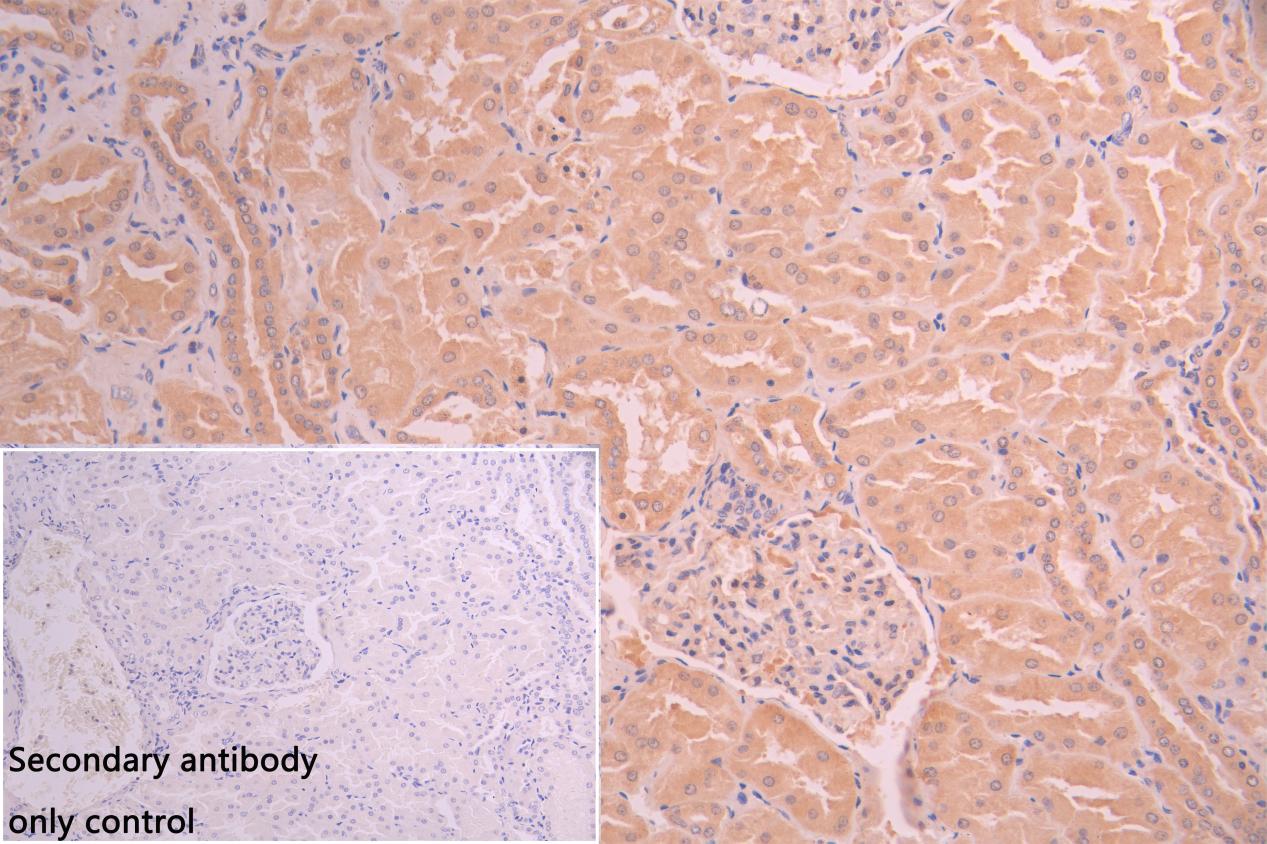
IHC image of CSB-PA882123LA01HU diluted at 1:50 and staining in paraffin-embedded human kidney tissue performed on a Leica BondTM system. After dewaxing and hydration, antigen retrieval was mediated by high pressure in a citrate buffer (pH 6.0). Section was blocked with 10% normal goat serum 30min at RT. Then primary antibody (1% BSA) was incubated at 4C overnight. The primary is detected by a Goat anti-rabbit polymer IgG labeled by HRP and visualized using 0.05% DAB. Secondary antibody only control: uses 1% BSA instead of primary antibody |
|
|
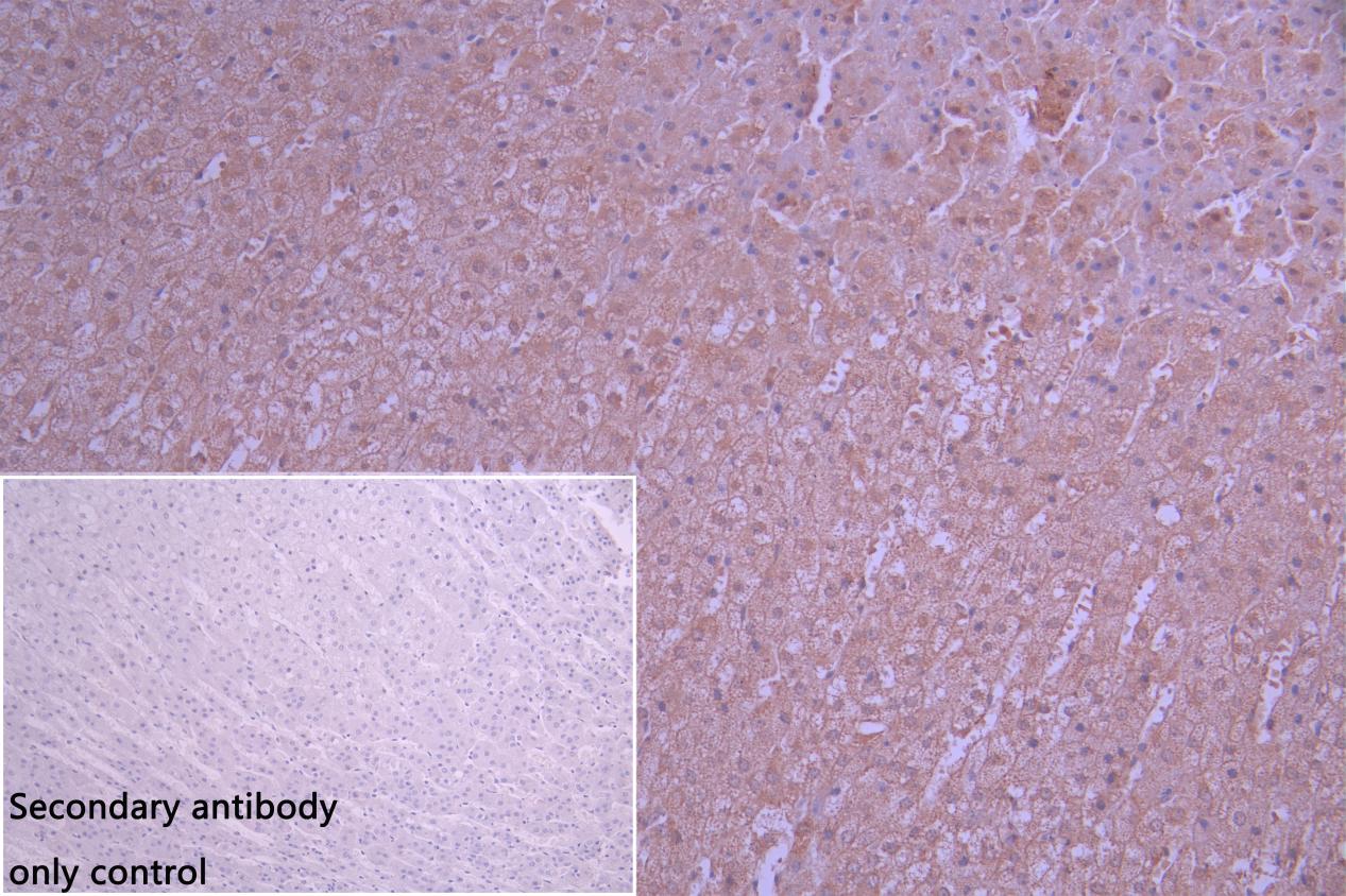
IHC image of CSB-PA882123LA01HU diluted at 1:50 and staining in paraffin-embedded human adrenal gland tissue performed on a Leica BondTM system. After dewaxing and hydration, antigen retrieval was mediated by high pressure in a citrate buffer (pH 6.0). Section was blocked with 10% normal goat serum 30min at RT. Then primary antibody (1% BSA) was incubated at 4C overnight. The primary is detected by a Goat anti-rabbit polymer IgG labeled by HRP and visualized using 0.05% DAB. Secondary antibody only control: uses 1% BSA instead of primary antibody |
|
|
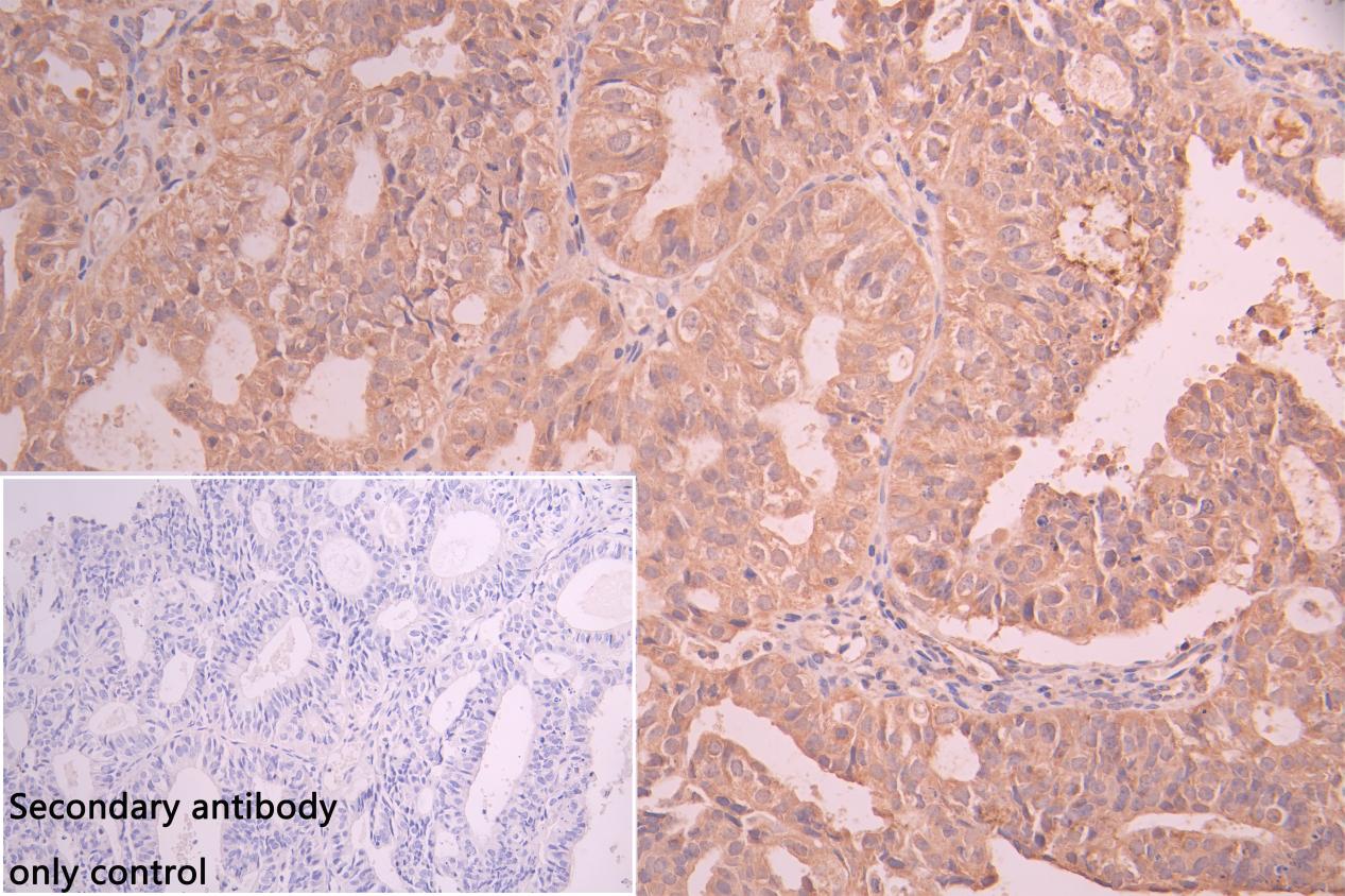
IHC image of CSB-PA882123LA01HU diluted at 1:50 and staining in paraffin-embedded human ovarian cancer performed on a Leica BondTM system. After dewaxing and hydration, antigen retrieval was mediated by high pressure in a citrate buffer (pH 6.0). Section was blocked with 10% normal goat serum 30min at RT. Then primary antibody (1% BSA) was incubated at 4C overnight. The primary is detected by a Goat anti-rabbit polymer IgG labeled by HRP and visualized using 0.05% DAB. Secondary antibody only control: uses 1% BSA instead of primary antibody |
|
|
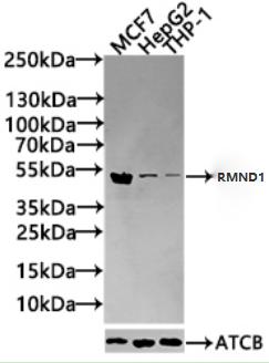
Western Blot Positive WB detected in: MCF7 whole cell lysate(30µg), HepG2 whole cell lysate(30µg), THP-1 whole cell lysate(30µg) All lanes: RMND1 antibody at 1:1000 Secondary Goat polyclonal to rabbit IgG at 1/20000 dilution Predicted band size: 52,28,24 kDa Observed band size: 52 kDa Exposure time: 120s |
Produktgarantie und fachkundiger Support
