
ATF2 Recombinant Monoclonal Antibody, Clone: [3D12], Unconjugated, Rabbit
Artikelnummer:
CSB-RA002270A0HU
- Bilder (2)
| Artikelname: | ATF2 Recombinant Monoclonal Antibody, Clone: [3D12], Unconjugated, Rabbit |
| Artikelnummer: | CSB-RA002270A0HU |
| Hersteller Artikelnummer: | CSB-RA002270A0HU |
| Alternativnummer: | CSB-RA002270A0HU-100UL, CSB-RA002270A0HU-50UL |
| Hersteller: | Cusabio |
| Wirt: | Rabbit |
| Kategorie: | Antikörper |
| Applikation: | ELISA, IHC, WB |
| Spezies Reaktivität: | Human |
| Konjugation: | Unconjugated |
| Alternative Synonym: | Cyclic AMP-dependent transcription factor ATF-2, Activating transcription factor 2, Cyclic AMP-responsive element-binding protein 2, CREB-2, cAMP-responsive element-binding protein 2, HB16, Histone acetyltransferase ATF2, cAMP response element-binding protein CRE-BP1, ATF2, CREB2, CREBP1 |
| Klonalität: | Monoclonal |
| Klon-Bezeichnung: | [3D12] |
| UniProt: | P15336 |
| Puffer: | Rabbit IgG in 10mM phosphate buffered saline , pH 7.4, 150mM sodium chloride, 0.05% BSA, 0.02% sodium azide and 50% glycerol. |
| Reinheit: | Affinity-chromatography |
| Formulierung: | Liquid |
| Target-Kategorie: | ATF2 |
| Antibody Type: | Recombinant Antibody |
| Application Verdünnung: | Recommended dilution: WB:1:500-1:5000, IHC:1:50-1:200 |
|
|
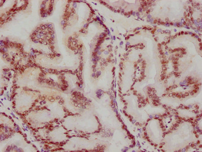
IHC image of CSB-RA002270A0HU diluted at 1:115.5 and staining in paraffin-embedded human prostate tissue performed on a Leica BondTM system. After dewaxing and hydration, antigen retrieval was mediated by high pressure in a citrate buffer (pH 6.0) . Section was blocked with 10% normal goat serum 30min at RT. Then primary antibody (1% BSA) was incubated at 4°C overnight. The primary is detected by a biotinylated secondary antibody and visualized using an HRP conjugated SP system. |
|
|
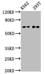
Western Blot Positive WB detected in: K562 whole cell lysate, 293T whole cell lysate All lanes: ATF2 antibody at 1.2µg/ml Secondary Goat polyclonal to rabbit IgG at 1/50000 dilution Predicted band size: 55, 36, 24, 49, 53, 14, 25, 16 KDa Observed band size: 70 KDa |
Produktgarantie und fachkundiger Support
