
ENO1 Recombinant Monoclonal Antibody, Clone: [8H9G12], Unconjugated
Artikelnummer:
CSB-RA007670MA1HU
- Bilder (6)
| Artikelname: | ENO1 Recombinant Monoclonal Antibody, Clone: [8H9G12], Unconjugated |
| Artikelnummer: | CSB-RA007670MA1HU |
| Hersteller Artikelnummer: | CSB-RA007670MA1HU |
| Alternativnummer: | CSB-RA007670MA1HU-100UL, CSB-RA007670MA1HU-50UL |
| Hersteller: | Cusabio |
| Kategorie: | Antikörper |
| Applikation: | ELISA, FC, IF, IHC, WB |
| Spezies Reaktivität: | Human, Mouse, Rat |
| Konjugation: | Unconjugated |
| Alternative Synonym: | 2 phospho D glycerate hydro lyase antibody, 2-phospho-D-glycerate hydro-lyase antibody, Alpha enolase antibody, Alpha enolase like 1 antibody, Alpha-enolase antibody, C myc promoter binding protein antibody, C-myc promoter-binding protein antibody, EC 4.2.1.11 antibody, eno1 antibody, ENO1L1 antibody, ENOA_HUMAN antibody, Enolase 1 (alpha) antibody, Enolase 1 (alpha) like 1 antibody, Enolase 1 antibody, Enolase alpha antibody, MBP 1 antibody, MBP-1 antibody, MBP1 antibody, MBPB1 antibody, MPB 1 antibody, MPB-1 antibody, MPB1 antibody, MYC promoter binding protein 1 antibody, NNE antibody, Non neural enolase antibody, Non-neural enolase antibody, Phosphopyruvate hydratase antibody, Plasminogen binding protein antibody, Plasminogen-binding protein antibody, PPH antibody, Tau crystallin antibody |
| Klonalität: | Monoclonal |
| Klon-Bezeichnung: | [8H9G12] |
| UniProt: | P06733 |
| Puffer: | Preservative: 0.03% Proclin 300<br />Constituents: 50% Glycerol, 0.01M PBS, PH 7.4 |
| Reinheit: | Affinity-chromatography |
| Formulierung: | Liquid |
| Target-Kategorie: | ENO1 |
| Antibody Type: | Recombinant Antibody |
| Application Verdünnung: | Recommended dilution: WB:1:500-1:2000, IHC:1:50-1:200, IF:1:50-1:200, FC:1:50-1:200 |
|
|
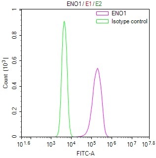
Overlay Peak curve showing Hela cells stained with CSB-RA007670MA1HU (red line) at 1:100. The cells were fixed in 4% formaldehyde and permeated by 0.2% TritonX-100 for10min. Then 10% normal goat serum to block non-specific protein-protein interactions followed by the antibody (1ug/1*106cells) for 45min at 4°C. The secondary antibody used was Fluorescein (FITC) AffiniPure Goat Anti-Human IgG, Fcgamma fragment specific at 1:200 dilution for 35min at 4°C.Control antibody (green line) was human IgG1 (1ug/1*106cells) used under the same conditions. Acquisition of >10,000 events was performed. |
|
|
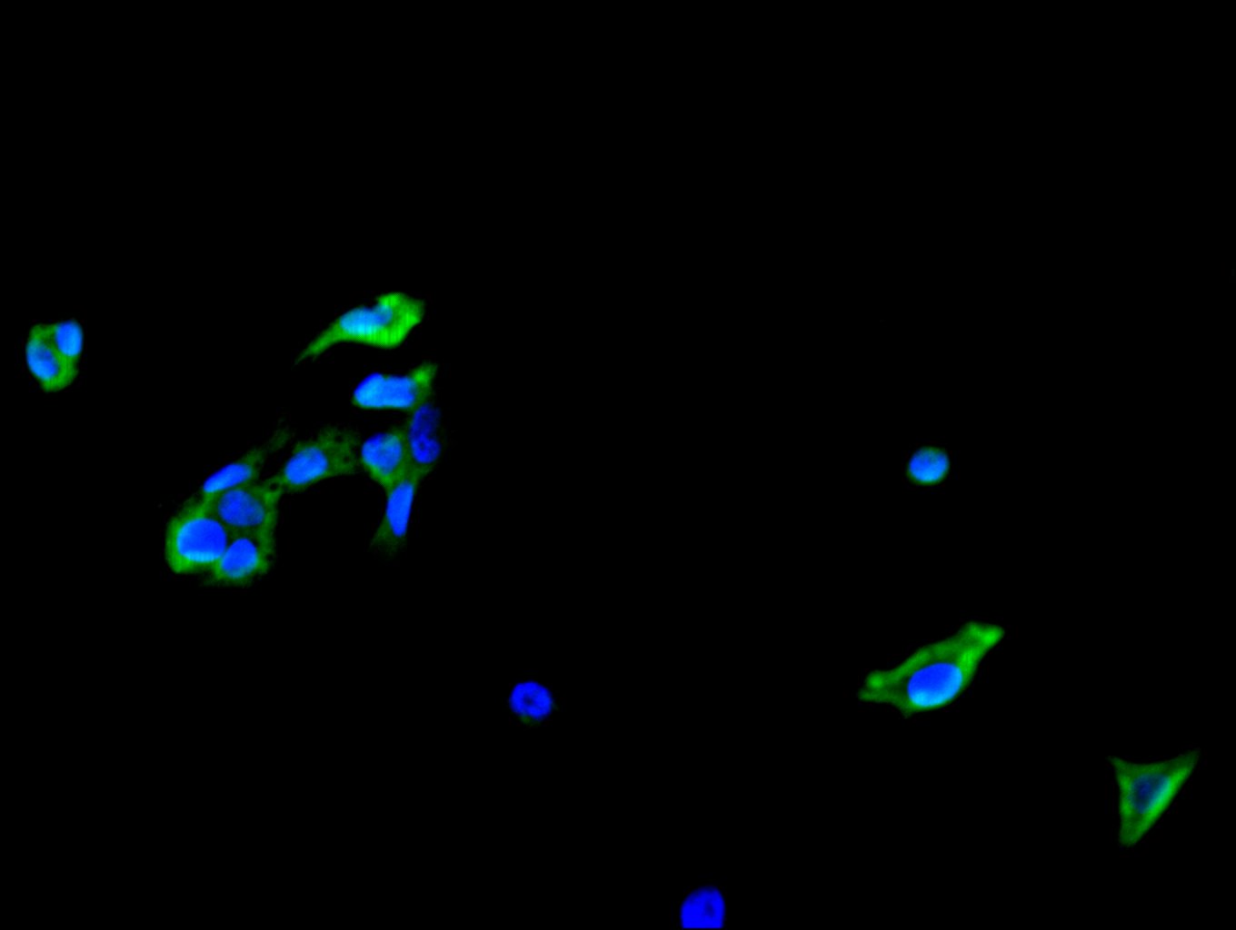
Immunofluorescence staining of HepG2 cell with CSB-RA007670MA1HU at 1:30 counter-stained with DAPI. The cells were fixed in 4% formaldehyde, permeabilized using 0.2% Triton X-100 and blocked in 10% normal Goat Serum. The cells were then incubated with the antibody overnight at 4C. The secondary antibody was Fluorescein (FITC) AffiniPure Goat Anti-Human IgG, Fcgamma fragment specific. |
|
|
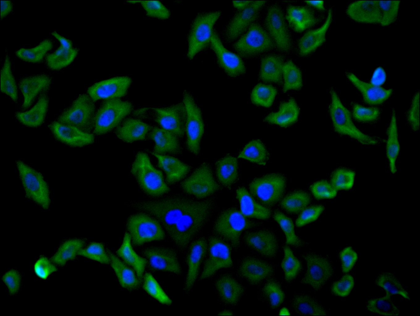
Immunofluorescence staining of MCF-7 cell with CSB-RA007670MA1HU at 1:30 counter-stained with DAPI. The cells were fixed in 4% formaldehyde, permeabilized using 0.2% Triton X-100 and blocked in 10% normal Goat Serum. The cells were then incubated with the antibody overnight at 4C. The secondary antibody was Fluorescein (FITC) AffiniPure Goat Anti-Human IgG, Fcgamma fragment specific. |
|
|
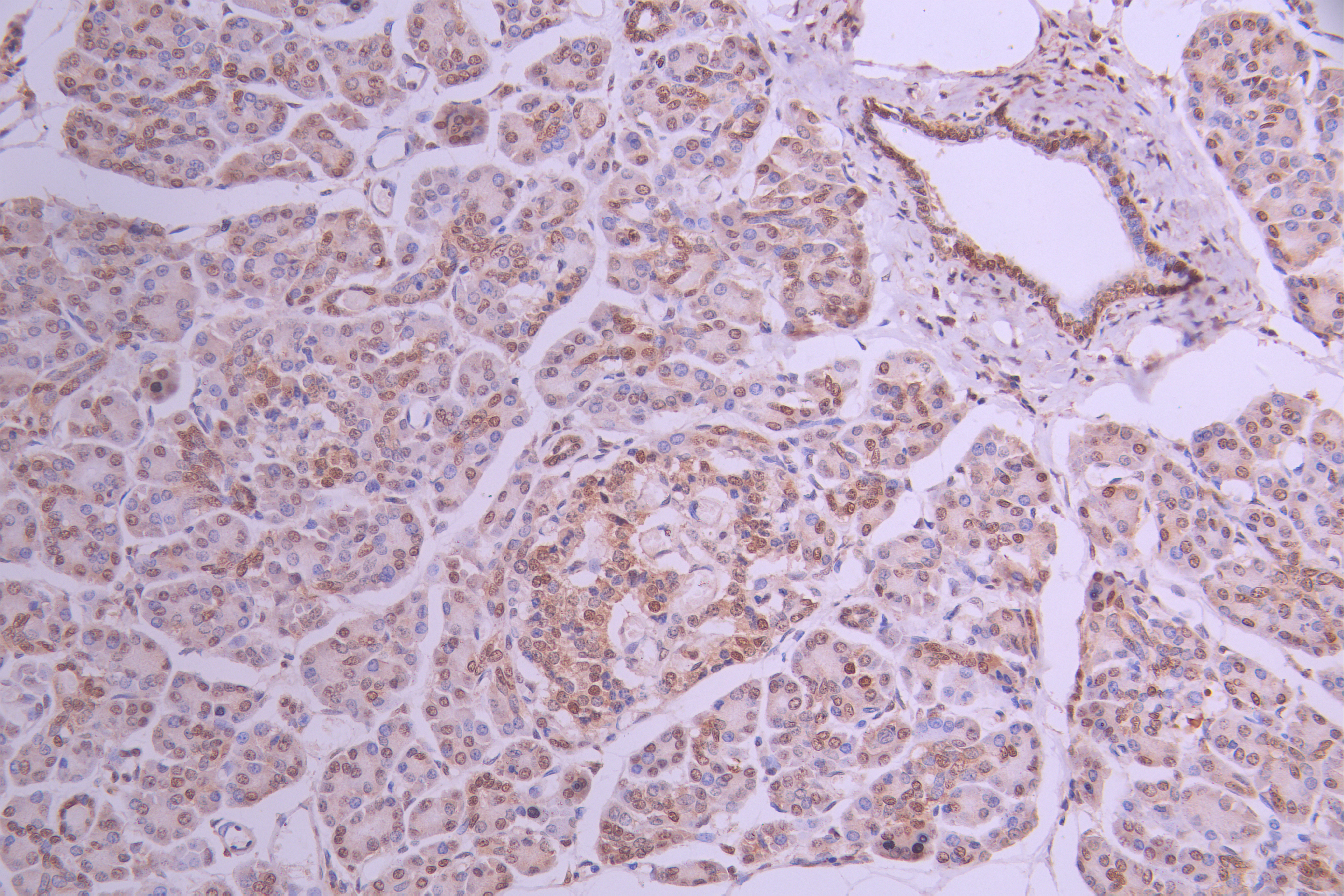
IHC image of CSB-RA007670MA1HU diluted at 1:50 and staining in paraffin-embedded human pancreatic tissue performed on a Leica BondTM system. After dewaxing and hydration, antigen retrieval was mediated by high pressure in a citrate buffer (pH 6.0) . Section was blocked with 10% normal goat serum 30min at RT. Then primary antibody (1% BSA) was incubated at 4C overnight. The primary is detected by a Goat anti-human polymer IgG labeled by HRP and visualized using 0.05% DAB. |
|
|
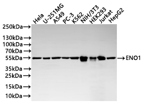
Western Blot Positive WB detected in: HeLa whole cell lysate(20µg) , U-251MG whole cell lysate(20µg) , A549 whole cell lysate(20µg) , PC-3 whole cell lysate(20µg) , K562 whole cell lysate(20µg) , NIH/3T3 whole cell lysate(20µg) , HEK293 whole cell lysate(20µg) , Jurkat whole cell lysate(20µg) , HepG2 whole cell lysate(20µg) All lanes: ENO1 antibody at 1:1000 Secondary Goat polyclonal to human IgG at 1/40000 dilution Predicted band size: 47 kDa Observed band size: 55 kDa Exposure time:5s |
|
|
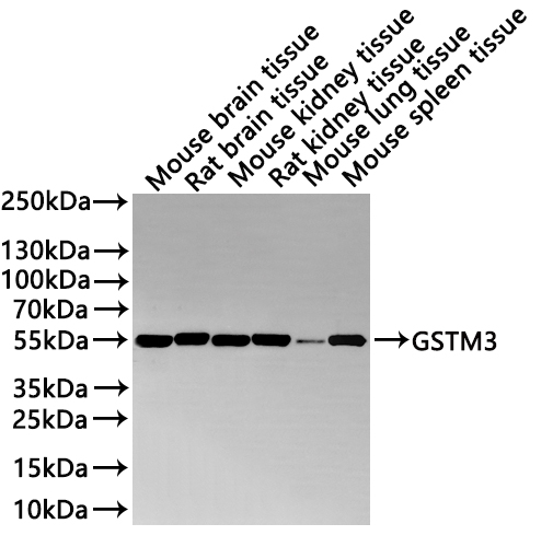
Western Blot Positive WB detected in: Mouse brain tissue lysate(20µg) , Rat brain tissue lysate(20µg) , Mouse kidney tissue lysate(20µg) , Rat kidney tissue lysate(20µg) , Mouse lung tissue lysate(20µg) , Mouse spleen tissue lysate(20µg) All lanes: ENO1 antibody at 1:1000 Secondary Goat polyclonal to human IgG at 1/40000 dilution Predicted band size: 47.2 kDa Observed band size: 55 kDa Exposure time: 2min |
Produktgarantie und fachkundiger Support
