
SDHA antibody [GT20710], IgG1, Unconjugated, Mouse, Monoclonal
Artikelnummer:
GTX632636
- Bilder (9)
| Artikelname: | SDHA antibody [GT20710], IgG1, Unconjugated, Mouse, Monoclonal |
| Artikelnummer: | GTX632636 |
| Hersteller Artikelnummer: | GTX632636 |
| Alternativnummer: | GTX632636-100,GTX632636-25 |
| Hersteller: | GeneTex |
| Wirt: | Mouse |
| Kategorie: | Antikörper |
| Applikation: | ICC, IHC-P, WB |
| Spezies Reaktivität: | Human, Mouse, Rat |
| Immunogen: | Recombinant protein encompassing a sequence within the N-terminus region of human SDHA. The exact sequence is proprietary. |
| Konjugation: | Unconjugated |
| Alternative Synonym: | succinate dehydrogenase complex flavoprotein subunit A , CMD1GG , FP , PGL5 , SDH1 , SDH2 , SDHF |
| Klonalität: | Monoclonal |
| Konzentration: | 1 mg/ml (Please refer to the vial label for the specific concentration.) |
| Klon-Bezeichnung: | [GT20710] |
| Molekulargewicht: | 73 |
| Isotyp: | IgG1 |
| NCBI: | 6389 |
| UniProt: | P31040 |
| Puffer: | 1XPBS pH7, 20% Glycerol, no Preservative. |
| Reinheit: | Affinity purified by Protein G. |
| Formulierung: | Liquid |
| Anwendungsbeschreibung: | WB: 1:500-1:3000. ICC/IF: 1:100-1:1000. IHC-P: 1:100-1:1000. *Optimal dilutions/concentrations should be determined by the researcher.Not tested in other applications. |
|
|
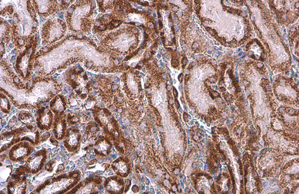
GTX632636 IHC-P Image |
|
|
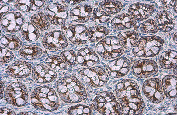
SDHA antibody [GT20710] detects SDHA protein at mitochondria by immunohistochemical analysis.Sample: Paraffin-embedded mouse liver.SDHA stained by SDHA antibody [GT20710] (GTX632636) diluted at 1:200.Antigen Retrieval: Citrate buffer, pH 6.0, 15 min |
|
|
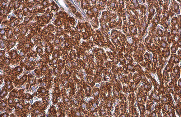
SDHA antibody [GT20710] detects SDHA protein at mitochondria by immunohistochemical analysis.Sample: Paraffin-embedded rat colon.SDHA stained by SDHA antibody [GT20710] (GTX632636) diluted at 1:200.Antigen Retrieval: Citrate buffer, pH 6.0, 15 min |
|
|
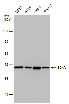
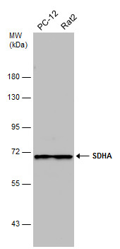
Various whole cell extracts (30 μg) were separated by 7.5% SDS-PAGE, and the membrane was blotted with SDHA antibody [GT20710] (GTX632636) diluted at 1:3000. |
|
|
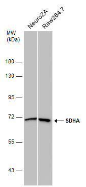
Various whole cell extracts (30 μg) were separated by 7.5% SDS-PAGE, and the membrane was blotted with SDHA antibody [GT20710] (GTX632636) diluted at 1:3000. |
|
|
Various whole cell extracts (30 μg) were separated by 7.5% SDS-PAGE, and the membrane was blotted with SDHA antibody [GT20710] (GTX632636) diluted at 1:3000. |
|
|
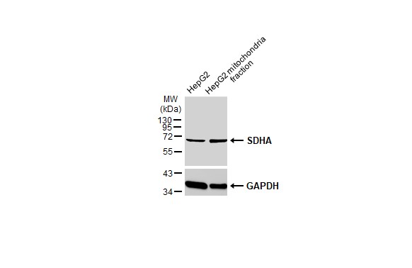
HepG2 and mitochondria extracts (30 μg) were separated by SDS-PAGE, and the membrane was blotted with SDHA antibody [GT20710] (GTX632636) diluted at 1:1000. The HRP-conjugated anti-mouse IgG antibody (GTX213111-01) was used to detect the primary antibody. |
|
|
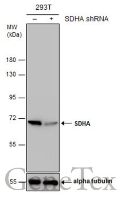
Non-transfected (–) and transfected (+) 293T whole cell extracts (30 μg) were separated by 7.5% SDS-PAGE, and the membrane was blotted with SDHA antibody [GT20710] (GTX632636) diluted at 1:1000. |
|
|
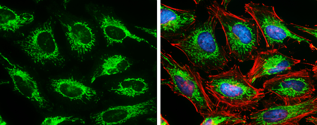
SDHA antibody [GT20710] detects SDHA protein at mitochondria by immunofluorescent analysis. Sample: HeLa cells were fixed in 4% paraformaldehyde at RT for 15 min. Green: SDHA protein stained by SDHA antibody [GT20710] (GTX632636) diluted at 1:500. Red: Phalloidin, a cytoskeleton marker, diluted at 1:100. Blue: Hoechst 33342 staining. |
Produktgarantie und fachkundiger Support
