
CTCF Antibody, Unconjugated, Rabbit, Polyclonal
Artikelnummer:
PRS-14-881
- Bilder (8)
| Artikelname: | CTCF Antibody, Unconjugated, Rabbit, Polyclonal |
| Artikelnummer: | PRS-14-881 |
| Hersteller Artikelnummer: | 14-881 |
| Alternativnummer: | PRS-14-881-100 |
| Hersteller: | ProSci |
| Wirt: | Rabbit |
| Kategorie: | Antikörper |
| Applikation: | ChIP, IF, IHC, IP, WB |
| Spezies Reaktivität: | Human, Mouse, Rat |
| Immunogen: | Recombinant fusion protein containing a sequence corresponding to amino acids 1-260 of human CTCF (NP_006556.1). |
| Konjugation: | Unconjugated |
| Alternative Synonym: | CTCF, MRD21 |
| Application Verdünnung: | Optimal dilutions for each application to be determined by the researcher. |
| Anwendungsbeschreibung: | WB: 1:1000 - 1:2000IHC: 1:50 - 1:200IF: 1:50 - 1:200IP: 1:50 - 1:100ChIP: 1:50 - 1:200 |
|
|
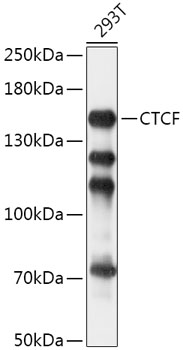
Western blot analysis of extracts of 293T cells, using CTCF antibody (14-881) at 1:1000 dilution. Secondary antibody: HRP Goat Anti-Rabbit IgG (H+L) at 1:10000 dilution. Lysates/proteins: 25ug per lane. Blocking buffer: 3% nonfat dry milk in TBST. Detection: ECL Basic Kit. Exposure time: 5s. |
|
|
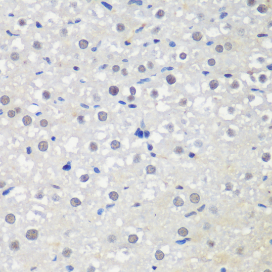
Immunohistochemistry of paraffin-embedded rat liver using CTCF antibody (14-881) at dilution of 1:200 (40x lens). |
|
|
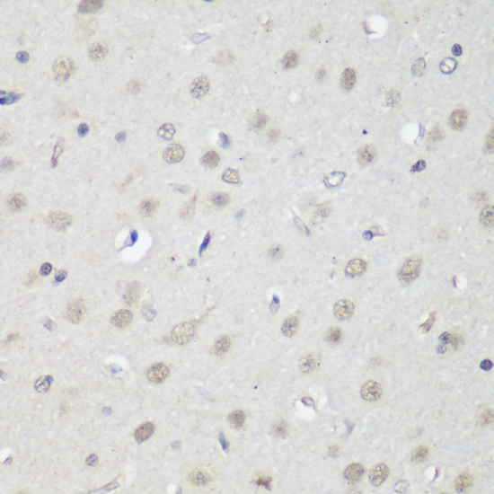
Immunohistochemistry of paraffin-embedded rat brain using CTCF antibody (14-881) at dilution of 1:200 (40x lens). |
|
|
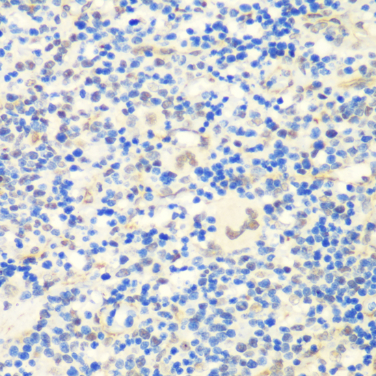
Immunohistochemistry of paraffin-embedded rat spleen using CTCF antibody (14-881) at dilution of 1:200 (40x lens). |
|
|
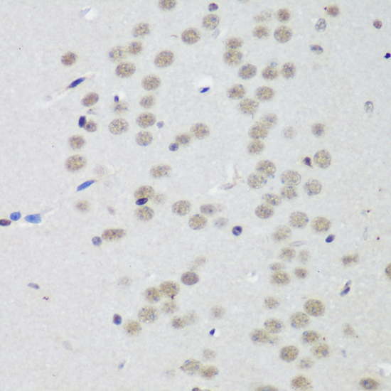
Immunohistochemistry of paraffin-embedded mouse brain using CTCF antibody (14-881) at dilution of 1:200 (40x lens). |
|
|
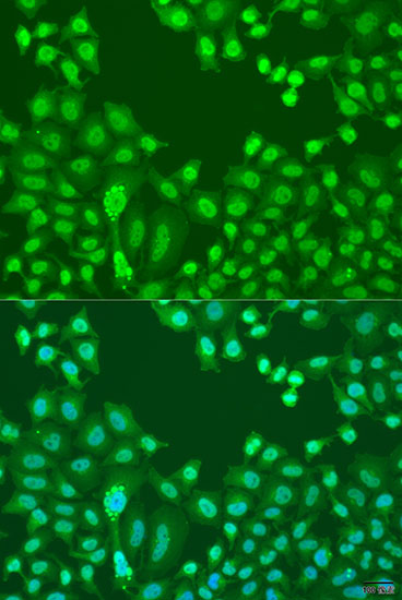
Immunofluorescence analysis of U2OS cells using CTCF antibody (14-881) at dilution of 1:100. Blue: DAPI for nuclear staining. |
|
|
Immunoprecipitation analysis of 200ug extracts of HeLa cells, using 3 ug CTCF antibody (14-881). Western blot was performed from the immunoprecipitate using CTCF antibody (14-881) at a dilition of 1:1000. |
|
|
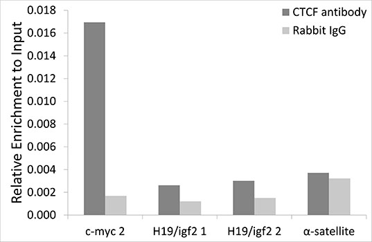
Chromatin immunoprecipitation analysis of extracts of HCT116 cells, using CTCF antibody (14-881) and rabbit IgG. The amount of immunoprecipitated DNA was checked by quantitative PCR. Histogram was constructed by the ratios of the immunoprecipitated DNA to the input. |
Produktgarantie und fachkundiger Support
