
Acetyl-Histone H3-K9 pAb, Unconjugated, Rabbit, Polyclonal
Artikelnummer:
PRS-22-814
- Bilder (8)

| Artikelname: | Acetyl-Histone H3-K9 pAb, Unconjugated, Rabbit, Polyclonal |
| Artikelnummer: | PRS-22-814 |
| Hersteller Artikelnummer: | 22-814 |
| Alternativnummer: | PRS-22-814-100 |
| Hersteller: | ProSci |
| Wirt: | Rabbit |
| Kategorie: | Antikörper |
| Applikation: | ChIP, IF, IHC, IP, WB |
| Spezies Reaktivität: | Mouse, Rat |
| Immunogen: | A synthetic peptide of human Acetyl-Histone H3-K9 |
| Konjugation: | Unconjugated |
| Alternative Synonym: | H3F3A, H3t, H3.4, H3/g, H3FT |
| Application Verdünnung: | Optimal dilutions for each application to be determined by the researcher. |
| Anwendungsbeschreibung: | WB: 1:500 - 1:2000IHC: 1:50 - 1:200IF: 1:50 - 1:200IP: 1:50 - 1:200ChIP: 1:20 - 1:100CHIP seq: 1:20 - 1:50 |
|
|
Immunofluorescence analysis of NIH/3T3 cells using Acetyl-Histone H3-K9 antibody (22-814) at dilution of 1:100 (40x lens). NIH/3T3 cells were treated by TSA (1 uM) at 37°C for 18 hours (left). Blue: DAPI for nuclear staining. |
|
|
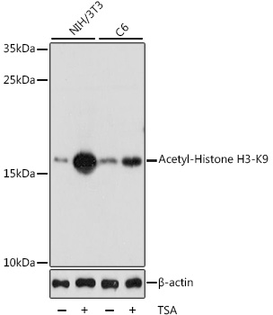
Western blot analysis of extracts of various cell lines, using Acetyl-Histone H3-K9 antibody (22-814) at 1:1000 dilution. Both NIH/3T3 cells and C6 cells were treated by TSA (1 uM) at 37°C for 18 hours. Secondary antibody: HRP Goat Anti-Rabbit IgG (H+L) at 1:10000 dilution. Lysates/proteins: 25ug per lane. Blocking buffer: 3% nonfat dry milk in TBST. Detection: ECL Basic Kit. Exposure time: 1s. |
|
|
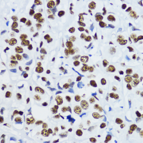
Immunohistochemistry of paraffin-embedded human mammary cancer using Acetyl-Histone H3-K9 antibody (22-814) at dilution of 1:200 (40x lens). |
|
|
Immunohistochemistry of paraffin-embedded human colon using Acetyl-Histone H3-K9 antibody (22-814) at dilution of 1:200 (40x lens). |
|
|
Immunohistochemistry of paraffin-embedded rat ovary using Acetyl-Histone H3-K9 antibody (22-814) at dilution of 1:200 (40x lens). |
|
|
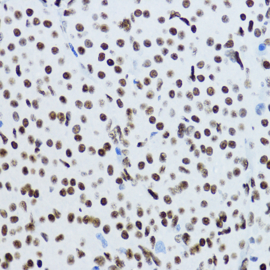
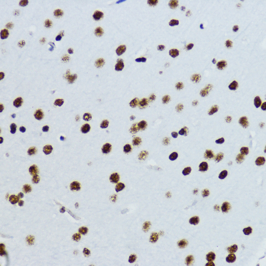
Immunohistochemistry of paraffin-embedded mouse brain using Acetyl-Histone H3-K9 antibody (22-814) at dilution of 1:200 (40x lens). |
|
|
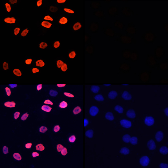
Immunofluorescence analysis of HeLa cells using Acetyl-Histone H3-K9 antibody (22-814) at dilution of 1:100 (40x lens). HeLa cells were treated by TSA (1 uM) at 37°C for 18 hours (left). Blue: DAPI for nuclear staining. |
|
|
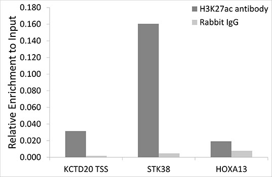
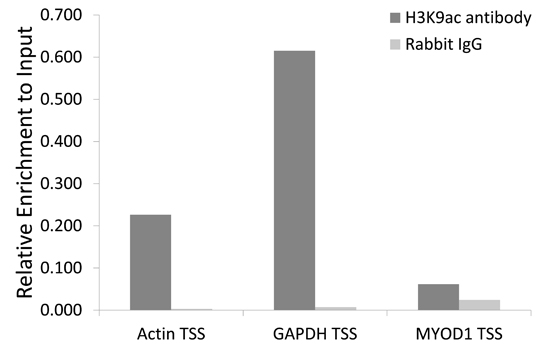
Chromatin immunoprecipitation analysis of extracts of 293 cell line, using H3K9ac antibody (22-814) and rabbit IgG. The amount of immunoprecipitated DNA was checked by quantitative PCR. Histogram was constructed by the ratios of the immunoprecipitated DNA to the input. |
Produktgarantie und fachkundiger Support
