Anti-PAX6 antibody has been tested by ELISA and Immunofluorescence and suitable for Western Blotting. Specific conditions for reactivity should be optimized by the end user. Expect a band approximately 46.7 kDa in size corresponding to PAX6 by western bl
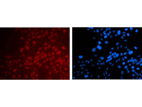
Immunofluorescence Microscopy of Mouse anti-PAX6 antibody. Tissue: Hela Cell,20X. Fixation: 4% PFA room temp for 10 min. Antigen retrieval: not required. Primary antibody: PAX6 antibody at 1:100 for 1 h at RT. Secondary antibody: goat anti-mouse secondary antibody at 1:1,000 for 45 min at RT. Localization: PAX6 is nuclear. Staining: PAX6 as red fluorescent signal with DAPI (blue) nuclear counterstain. Images courtesy of Yang Xiang with Boston Childrens Hospital.
* Mehrwertsteuer und Versandkosten nicht enthalten. Irrtümer und Preisänderungen vorbehalten