Anti-GFP / Green Fluorescent Protein Polyclonal Antibody, Rabbit
Biozol Catalog Number:
ABI-30-1330
Supplier Catalog Number:
30-1330
Alternative Catalog Number:
ABI-30-1330-0.1MG
Manufacturer:
Abeomics
Host:
Rabbit
Category:
Antikörper
Application:
ICC, IP, WB
Immunogen:
EGFP, a native full-length protein
Green fluorescence protein (GFP) is a 27 KDa protein derived from the bioluminiscent jellyfish Aquorea victoria, emiting green light (lambda=509 nm) when excited (excitation by Blue or UV light, absorption peak lambda=395 nm). GFP is a useful tool in cell biology research, as its intrinsic fluorescence can be visualized in living cells. Light-stimulated GFP fluorescence is species-independent and a fluorescence has been reported from many different types of GFP-expressing hosts, including microbes, invertebrates, vertebrates and plants. No exogenous substrates and cofactors are required for the fluorescence of GFP, since GFP autocatalytically forms a fluorescent pigment from natural amino acids present in the nascent protein. GFP fluorescence is stable under fixation conditions and suitable for a variety of applications. GFP is widely used as a reporter (tag) for gene expression, enabling researchers to visualize and localize GFP-tagged proteins within living cells without any further staining. Other applications of GFP include measurement of distance between proteins through fluorescence energy transfer (FRET) protocols. To increase a fluorescence intensity of GFP, chomophore mutations have been created. The EnhancedGFP has a fluorescence 35 times more intense than the wt-GFP. Mutagenesis of GFP has produced also many mutants (e.g. Yellow Fluorescent Protein, Cyan Fluorescent Protein) with warying spectral properties. Antibodies raised against full-length GFP variants should also detect other variants of the protein.
Clonality:
Polyclonal
Purity:
Purified from rabbit serum by affinity chromatography
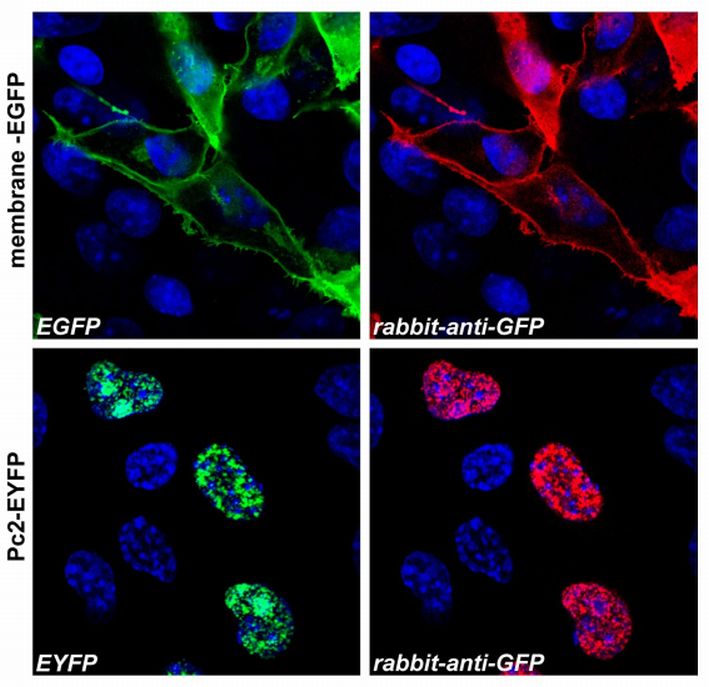
Figure 1: Confocal microscopy images of COS-7 cells transfected with expression constructs encoding membrane-tethered EGFP (membrane-EGFP, top) or nuclear Polycomb 2-EYFP fusion protein (Pc2-EYFP, bottom). The natural fluorescence of the produced proteins is shown in the green channel (left), polyclonal anti-GFP antibody signal was detected in the red channel (right). The system was carefully tested for overlap of these two optical channels and images were scanned separately in sequential scanni
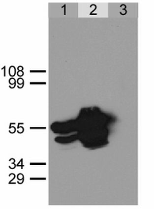
Figure 2: Immunoprecipitation of GFP-NLS from HEK293 cells using anti-GFP antibody.HEK293 cells were transfected with expression construct encoding GFP-NLS protein. Twenty hours post transfection cells were lysed in non-denaturating conditions (Lysis buffer: 20 mM Tris, pH 7.5, 100 mM NaCl, 0.5% Triton X-100, inhibitors of proteases). Aliquots of cell lysate were immunoprecipitated using a polyclonal anti-GFP antibody (lane 2) or a pre-immune rabbit serum (lane 3). Immunoprecipitates together w
* VAT and and shipping costs not included. Errors and price changes excepted