0.02 M Potassium Phosphate, 0.15 M Sodium Chloride, pH 7.2
Form:
Lyophilized
Target:
Goat
Antibody Type:
Secondary Antibody
Application Dilute:
FLISA: >1:20,000, IF Microscopy: >1:5,000, WB: >1:10,000
Application Notes:
Anti-Goat IgG Antibody ATTO 594 Conjugated has been tested by dot blot and western blot and is designed for STED microscopy, FRET, immunofluorescence microscopy, fluorescence based plate assays (FLISA) and fluorescent western blotting. This product is al
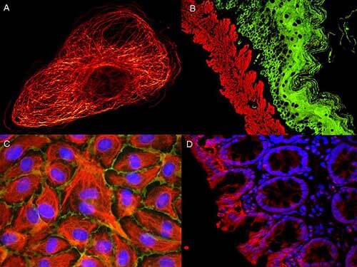
ATTO dyes can be used for multicolor immunofluorescent detection with low background and high signal. Examples shown are: A. Tubulin in PtK2- male Rat Kangaroo Kidney Epithelial Cells was detected using ATTO 532 labeled secondary antibody. B. Muscle alpha-actin was stained with a mouse primary antibody and ATTO 488 anti-mouse IgG (green) while Cytokeratin was stained with polyclonal rabbit anti-cytokeratin and ATTO 647N anti-rabbit IgG (red). C. HUVEC (Human umbilical vein endothelial cells were stained with anti- Vimentin-ATTO 532 (green), anti-E-Cadherin-ATTO 655 (red) and DAPI (blue). D. Rat colon sections were stained with Anti-Aquaporin 3-ATTO 594 antibody. Hoechst 33342 (blue) is used as counterstain. Images provided courtesy of Dr. Jörg Reichwein, ATTO-TEC GmbH
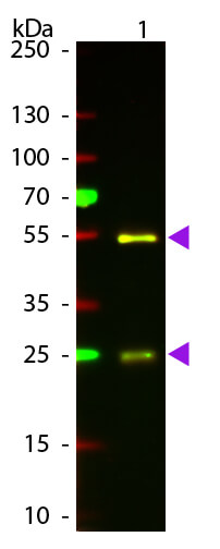
Western Blot of Atto 594 Conjugated Rabbit Anti-Goat IgG Pre-Absorbed Secondary Antibody. Lane 1: Goat IgG. Lane 2: None. Load: 50 ng per lane. Primary antibody: None. Secondary antibody: Atto 594 rabbit secondary antibody at 1:1,000 in MB-070 for 60 min at RT. Block: MB-070 for 30 min at RT. Predicted/Observed size: 28 & 55 kDa, 28 & 55 kDa for Goat IgG. Other band(s): None.
* VAT and and shipping costs not included. Errors and price changes excepted