![]()
|
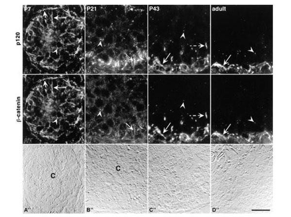
Colocalization of p120 and beta-catenin using Anti-Mouse IgG1 FITC (p/n 610-4240) during postnatal testis development. Here, the 8D11 antibody was used to immunostain for p120. In all locations, p120 and beta-catenin showed exact colocalization. At Postnatal Day 7 (A), diffuse immunostaining outlined all cells of the developing epithelium (solid arrow). In addition, punctate structures (arrowhead) were observed throughout the tubule. At the tubule periphery, intense immunostaining was associated with peritubular cells (dashed arrows). From Day 21 through adulthood, p120 and beta-catenin colocalized at basal inter-Sertoli junctions (arrows inB-D), and at punctate, spermatocyte-associated structures (arrowheads inB-D). In addition, extended, linear immunostaining at the level of spermatocytes was observed at Day 43 (CandC', dashed arrows). Corresponding DIC images are shown inA'',B'',C'', andD''. The center (C) of Day 7 and Day 21 seminiferous tubules is indicated in the DIC images. In all images, the seminiferous tubule basement membrane is located at the bottom, and inC''andD'', the entire seminiferous epithelium is shown. Bar = 20 µm. Fig 2. PMID: 11906917. |
![]()
|
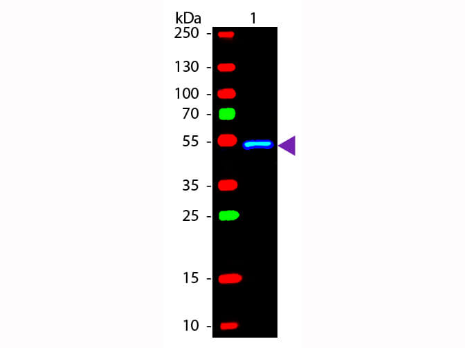
Western blot of Fluorescein conjugated Rabbit Anti-Mouse IgG1 (Gamma 1 chain) secondary antibody. Lane 1: Mouse IgG1. Lane 2: None. Load: 50 ng per lane. Primary antibody: None. Secondary antibody: Fluorescein rabbit secondary antibody at 1:1,000 for 60 min at RT. Blocking: MB-070 for 30 min at RT. Predicted/Observed size: 55 kDa, 55 kDa for Mouse IgG1 (Gamma 1 chain). Other band(s): None. |
![]()
|
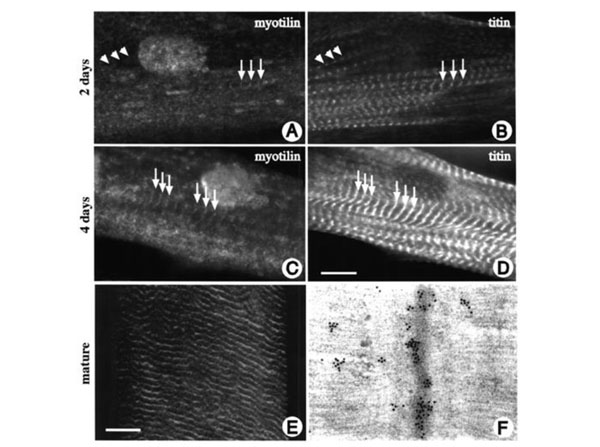
Localization of myotilin in differentiating and mature myocytes. Human skeletal muscle cells were grown on glass coverslips, differentiated for 4 days and stained with myotilin antibody (AandC) and with Z-disc specific titin antibody T12 mAb IgG1 (BandD) using Anti-Mouse IgG1 FITC. During the early stages of myofibril assembly, myotilin expression was faint and diffuse. The protein was concentrated at the areas where myofibrils are formed, and, although numerous Z-discs could be discerned, only very few of them contained myotilin (arrows in A and B). Only at the stage of myofibril alignment did myotilin appear at the mature, aligned Z-discs (arrows in C and D). In mature human skeletal muscle sections myotilin is found in a cross-striated pattern (E). Immunoelectron microscopic analysis of mature human skeletal muscle stained with myotilin antibody reveals a decoration of Z-discs (F). Bar=10µm. Fig 7. PMID: 12499399. |
![]()
|
Isolated hADSCs (IADSCs) were differentiated into SMCs using retinoic acid (RA), heparin was used as a positive control, while SK-UT-1 cells was used as a SMC control. Expression of SMC markers smooth muscle alpha actin (SM-alphaa, red, Texas Red Conjugated anti-Mouse IgG2, gamma2a chain specific, p/n 610-4941), desmin (green, Fluorescein Conjugated anti-Mouse IgG1, gamma1 chain specific, p/n 610-4240), smooth muscle myosin heavy chain (SM-MHC, red, Texas Red Conjugated anti-Mouse kappa, kappa chain specific, p/n 610-4910), and smoothelin (green, Fluorescein Conjugated anti-Mouse IgG1, gamma1 chain specific, p/n 610-4240) in differentiated SMCs was determined by indirect immunofluorescence. Nuclei were counter stained with DAPI (blue). Expression of all four markers can be seen in all the cells, particularly in RA differentiated SMCs. Fig. 5. PMID: 21373882. |