0.02 M Potassium Phosphate, 0.15 M Sodium Chloride, pH 7.2
Form:
Lyophilized
Target:
Mouse
Antibody Type:
Secondary Antibody
Application Dilute:
FLISA: >1:20,000, IF Microscopy: >1:5,000, WB: >1:10,000
Application Notes:
Anti-Mouse IgG (gamma 1, 2a, 2b and 3 chain) conjugated to ATTO 550 has been tested by western blot and is designed for STED microscopy, FRET, immunofluorescence microscopy, fluorescence based plate assays (FLISA) and fluorescent western blotting. This p
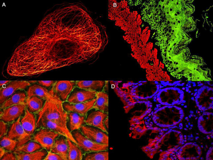
ATTO dyes can be used for multicolor immunofluorescent detection with low background and high signal. Examples shown are: A. Tubulin in PtK2- male Rat Kangaroo Kidney Epithelial Cells was detected using ATTO 532 labeled secondary antibody. B. Muscle alpha-actin was stained with a mouse primary antibody and ATTO 488 anti-mouse IgG (green) while Cytokeratin was stained with polyclonal rabbit anti-cytokeratin and ATTO 647N anti-rabbit IgG (red). C. HUVEC (Human umbilical vein endothelial cells were stained with anti- Vimentin-ATTO 532 (green), anti-E-Cadherin-ATTO 655 (red) and DAPI (blue). D. Rat colon sections were stained with Anti-Aquaporin 3-ATTO 594 antibody. Hoechst 33342 (blue) is used as counterstain. Images provided courtesy of Dr. Jörg Reichwein, ATTO-TEC GmbH
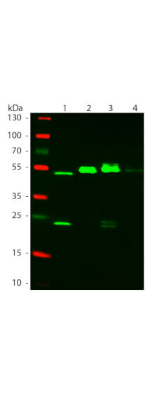
Western Blot of ATTO 550 Rabbit Anti-Mouse IgG (gamma 1, 2a, 2b, 3) secondary antibody. Lane 1: Mouse IgG1. Lane 2: Mouse IgG2a. Lane 3: Mouse IgG2b. Lane 4: Mouse IgG3. Load: 50 ng per lane. Primary antibody: None. Secondary antibody: ATTO 550 rabbit secondary antibody at 1:1,000 for 60 min at RT. Blocking: MB-070 for 30 min at RT. Predicted/Observed size: 25 & 55 kDa, 25 & 55 kDa for Mouse IgG Subclasses. Other band(s): None.
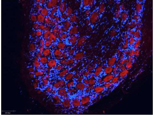
Atto(TM) dyes can be used for multicolor immunofluorescent detection with low background and high signal. Example shown here is Immunohistochemical staining using ATTO-550 Anti-Aquaporin 2-antibody (red) of paraffin embedded region of rat kidney showing a transversal cut of the inner medulla near to the renal papilla. Nuclei are visualized with Hoechst 33342 (blue). Images provided courtesy of Dr. Jörg Reichwein, ATTO-TEC GmbH
* VAT and and shipping costs not included. Errors and price changes excepted