0.02 M Potassium Phosphate, 0.15 M Sodium Chloride, pH 7.2
Form:
Lyophilized
Target:
Rabbit
Antibody Type:
Secondary Antibody
Application Dilute:
FLISA: >1:20,000, IF Microscopy: >1:5,000, WB: >1:10,000
Application Notes:
Anti-Rabbit IgG (H&L) conjugated to ATTO 425 has been tested by dot blot and western blot and is designed for STED microscopy, FRET, immunofluorescence microscopy, fluorescence based plate assays (FLISA) and fluorescent western blotting. This product is
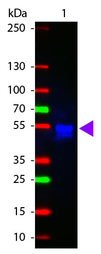
Western Blot of ATTO 425 conjugated Goat anti-Rabbit IgG antibody. Lane 1: Rabbit IgG. Lane 2: none. Load: 50 ng per lane. Primary antibody: none. Secondary antibody: ATTO 425 rabbit secondary antibody at 1:1,000 for 60 min at RT. Block: MB-070 for 30 min RT. Predicted/Observed size: 55 kDa, 28 kDa/55 kDa for Rabbit IgG. Other band(s): none.
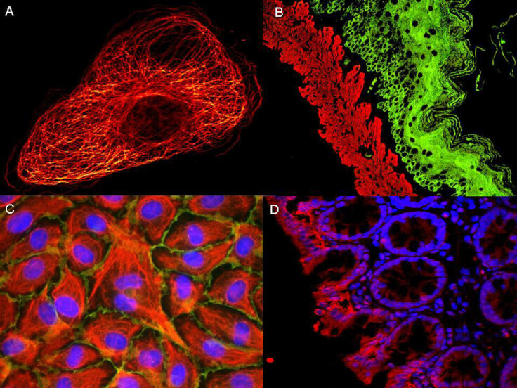
ATTO dyes can be used for multicolor immunofluorescent detection with low background and high signal. Examples shown are: A. Tubulin in PtK2- male Rat Kangaroo Kidney Epithelial Cells was detected using ATTO 532 labeled secondary antibody. B. Muscle alpha-actin was stained with a mouse primary antibody and ATTO 488 anti-mouse IgG (green) while Cytokeratin was stained with polyclonal rabbit anti-cytokeratin and ATTO 647N anti-rabbit IgG (red). C. HUVEC (Human umbilical vein endothelial cells were stained with anti- Vimentin-ATTO 532 (green), anti-E-Cadherin-ATTO 655 (red) and DAPI (blue). D. Rat colon sections were stained with Anti-Aquaporin 3-ATTO 594 antibody. Hoechst 33342 (blue) is used as counterstain. Images provided courtesy of Dr. Jörg Reichwein, ATTO-TEC GmbH
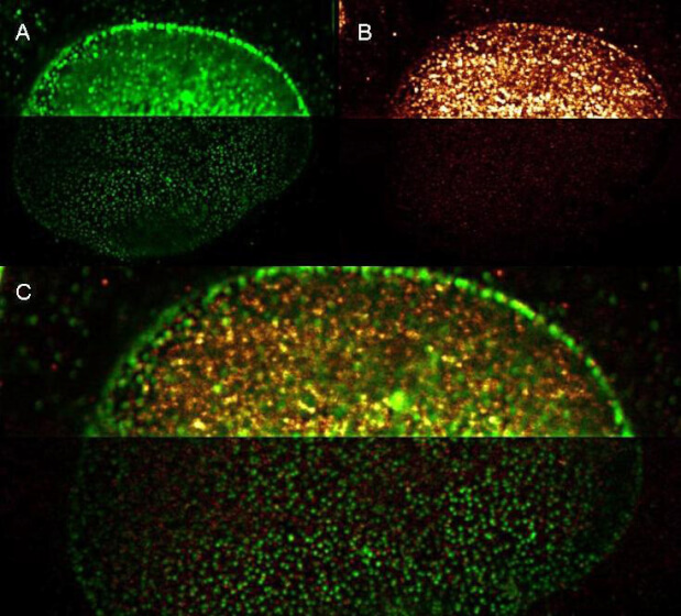
Rockland Dylight and ATTOdye conjugated antibodies provide high signal and low background for confocal microscopy (upper images) and high resolution Stimulated Emission Depletion (STED) Microscopy (lower images). Both Dylight and Atto conjugated secondary antibodies maintained robust, intense signal during repeated laser excitation and de-excitation used during STED microscopy. Shown here are: A. (Green) Mouse anti NuP (NuP=Nuclear Pore Protein) detected with Dylight 488 Goat anti mouse (610-141-121) B. (Red) Rabbit Anti Ezh1/2 Pab (Ezh=enhancer of zeste homology) with detection by Rockland ATTO 425 conjugated Goat anti Rabbit (611-151-122) (Red and Green) Images combined. Data was collected on a STED-CW TCS-SP5 Confocal system (Leica Microsystems) equipped with a DFC 350FX Camera allowing sequential acquisition in widefield, confocal and STED CW imaging modes and provided courtesy of: Myriam Gastard, PhD, personal communication, Leica Microsystems, Inc. USA
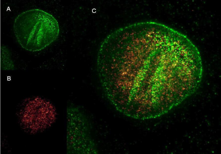
Rockland DyLight and ATTO dye conjugated antibodies provide high signal and low background for confocal microscopy and high resolution Stimulated Emission Depletion (STED) Microscopy. Both Dylight and Atto conjugated secondary antibodies maintained robust, intense signal during repeated laser excitation and de-excitation used during STED microscopy. Shown here are: A. (Green) Mouse anti NuP (NuP=Nuclear Pore Protein) detected with Dylight 488 Goat anti mouse (610-141-121) B. (Red) Rabbit Anti Ezh1/2 Pab (Ezh=enhancer of zeste homology) with detection by Rockland ATTO 425 conjugated Goat anti Rabbit (611-151-122) C. (Red and Green) Images combined. Data was collected on a STED-CW TCS-SP5 Confocal system (Leica Microsystems) equipped with a DFC 350FX Camera allowing sequential acquisition in wide-field, confocal and STED CW imaging modes and provided courtesy of: Myriam Gastard, PhD, personal communication, Leica Microsystems, Inc. USA
* VAT and and shipping costs not included. Errors and price changes excepted