
PIK3CA Rabbit pAb, Unconjugated, Polyclonal
Catalog Number:
ABB-A0265
- Images (9)


| Article Name: | PIK3CA Rabbit pAb, Unconjugated, Polyclonal |
| Biozol Catalog Number: | ABB-A0265 |
| Supplier Catalog Number: | A0265 |
| Alternative Catalog Number: | ABB-A0265-100UL,ABB-A0265-20UL,ABB-A0265-1000UL,ABB-A0265-500UL |
| Manufacturer: | ABclonal |
| Host: | Rabbit |
| Category: | Antikörper |
| Application: | ELISA, IF, IHC-P, WB |
| Species Reactivity: | Human |
| Immunogen: | Recombinant protein (or fragment).This information is considered to be commercially sensitive. |
| Conjugation: | Unconjugated |
| Alternative Names: | MCM, CCM4, CWS5, MCAP, PI3K, CLAPO, CLOVE, MCMTC, PI3K-alpha, p110-alpha, PIK3CA |
| Phosphatidylinositol 3-kinase is composed of an 85 kDa regulatory subunit and a 110 kDa catalytic subunit. The protein encoded by this gene represents the catalytic subunit, which uses ATP to phosphorylate PtdIns, PtdIns4P and PtdIns(4,5)P2. This gene has been found to be oncogenic and has been implicated in cervical cancers. A pseudogene of this gene has been defined on chromosome 22. |
| Clonality: | Polyclonal |
| Molecular Weight: | 124kDa |
| NCBI: | 5290 |
| UniProt: | P42336 |
| Purity: | Affinity purification |
| Sequence: | RLCLSICSVKGRKGAKEEHCPLAWGNINLFDYTDTLVSGKMALNLWPVPHGLEDLLNPIGVTGSNPNKETPCLELEFDWFSSVVKFPDMSVIEEHANWSVSREAGFSYSHAGLSNRLARDNELRENDKEQLKAISTRDPLSEITEQEKDFLWSHRHYCVTIPEILPKLLLSVKWNSRDEVAQMYCLVKDWPPIKPEQAME |
| Target: | PIK3CA |
| Antibody Type: | Primary Antibody |
| Application Dilute: | WB,1:5000 - 1:20000|IHC-P,1:50 - 1:200|IF/ICC,1:50 - 1:200|ELISA,Recommended starting concentration is 1 µg/mL. Please optimize the concentration based on your specific assay requirements. |
| Application Notes: | Cross-Reactivity: Human,Mouse,Rat. ResearchArea: Epigenetics Nuclear Signaling,Translation Control,Regulation of eIF4 and p70 S6 Kinase,Cancer,Signal Transduction,G protein signaling,Kinase,PI3K-Akt Signaling Pathway,mTOR Signaling Pathway,ErbB-HER Signaling Pathway,MAPK-Erk Signaling Pathway,MAPK-JNK Signaling Pathway,Cell Biology Developmental Biology,Apoptosis,Mitochondrial Control of Apoptosis,Inhibition of Apoptosis,Cell Adhesion,Cytoskeleton,Microtubules,Actins,TGF-b-Smad Signaling Pathway,ESC Pluripotency and Differentiation,Endocrine Metabolism,Lipid Metabolism,AMPK Signaling Pathway,Insulin Receptor Signaling Pathway,Warburg Effect,Immunology Inflammation,B Cell Receptor Signaling Pathway,T Cell Receptor Signaling Pathway,Jak-Stat-IL-6 Receptor Signaling Pathway,NF-kB Signaling Pathway,Toll-like Receptor Signaling Pathway,Cell Intrinsic Innate Immunity Signaling Pathway,TLR Signaling,Cardiovascular,Angiogenesis. Shipping: Ice Bag |
|
|
Immunohistochemistry analysis of paraffin-embedded Mouse kidney using PIK3CA Rabbit pAb (A0265) at dilution of 1:100 (40x lens). High pressure antigen retrieval performed with 0.01M Citrate buffer (pH 6.0) prior to IHC staining. |
|
|
Immunohistochemistry analysis of paraffin-embedded Rat liver using PIK3CA Rabbit pAb (A0265) at dilution of 1:100 (40x lens). High pressure antigen retrieval performed with 0.01M Citrate buffer (pH 6.0) prior to IHC staining. |
|
|
Western blot analysis of various lysates using PIK3CA Rabbit pAb (A0265) at 1:5000 dilution incubated overnight at 4°C. Secondary antibody: HRP-conjugated Goat anti-Rabbit IgG (H+L) (AS014) at 1:10000 dilution. Lysates/proteins: 25 µg per lane. Blocking buffer: 3% nonfat dry milk in TBST. Detection: ECL Basic Kit (RM00020). Exposure time: 20s. |
|
|
Immunohistochemistry analysis of paraffin-embedded Human breast cancer using PIK3CA Rabbit pAb (A0265) at dilution of 1:100 (40x lens). High pressure antigen retrieval performed with 0.01M Citrate buffer (pH 6.0) prior to IHC staining. |
|
|
Immunohistochemistry analysis of paraffin-embedded Mouse kidney using PIK3CA Rabbit pAb (A0265) at dilution of 1:100 (40x lens). High pressure antigen retrieval performed with 0.01M Citrate buffer (pH 6.0) prior to IHC staining. |
|
|
Immunohistochemistry analysis of paraffin-embedded Rat liver using PIK3CA Rabbit pAb (A0265) at dilution of 1:100 (40x lens). High pressure antigen retrieval performed with 0.01M Citrate buffer (pH 6.0) prior to IHC staining. |
|
|
Immunofluorescence analysis of NIH/3T3 cells using PIK3CA Rabbit pAb (A0265) at dilution of 1:100 (40x lens). Secondary antibody: Cy3-conjugated Goat anti-Rabbit IgG (H+L) (AS007) at 1:500 dilution. Blue: DAPI for nuclear staining. |
|
|
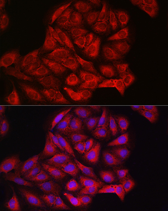
Immunofluorescence analysis of PC-12 cells using PIK3CA Rabbit pAb (A0265) at dilution of 1:100 (40x lens). Secondary antibody: Cy3-conjugated Goat anti-Rabbit IgG (H+L) (AS007) at 1:500 dilution. Blue: DAPI for nuclear staining. |
|
|
Immunofluorescence analysis of U2OS cells using PIK3CA Rabbit pAb (A0265) at dilution of 1:100 (40x lens). Secondary antibody: Cy3-conjugated Goat anti-Rabbit IgG (H+L) (AS007) at 1:500 dilution. Blue: DAPI for nuclear staining. |
Product Guarantee and Expert Support
