
CD68 Rabbit pAb, Unconjugated
Catalog Number:
ABB-A13286
- Images (6)


| Article Name: | CD68 Rabbit pAb, Unconjugated |
| Biozol Catalog Number: | ABB-A13286 |
| Supplier Catalog Number: | A13286 |
| Alternative Catalog Number: | ABB-A13286-100UL,ABB-A13286-20UL,ABB-A13286-1000UL,ABB-A13286-500UL |
| Manufacturer: | ABclonal |
| Host: | Rabbit |
| Category: | Antikörper |
| Application: | ELISA, IF, IHC-P, WB |
| Species Reactivity: | Human |
| Immunogen: | Synthetic peptide. This information is considered to be commercially sensitive. |
| Conjugation: | Unconjugated |
| Alternative Names: | GP110, LAMP4, SCARD1, CD68 |
| This gene encodes a 110-kD transmembrane glycoprotein that is highly expressed by human monocytes and tissue macrophages. It is a member of the lysosomal/endosomal-associated membrane glycoprotein (LAMP) family. The protein primarily localizes to lysosomes and endosomes with a smaller fraction circulating to the cell surface. It is a type I integral membrane protein with a heavily glycosylated extracellular domain and binds to tissue- and organ-specific lectins or selectins. The protein is also a member of the scavenger receptor family. Scavenger receptors typically function to clear cellular debris, promote phagocytosis, and mediate the recruitment and activation of macrophages. Alternative splicing results in multiple transcripts encoding different isoforms. |
| Clonality: | Polyclonal |
| Molecular Weight: | 37kDa |
| NCBI: | 968 |
| UniProt: | P34810 |
| Purity: | Affinity purification |
| Sequence: | NDCPHKKSATLLPSFTVTPTVTESTGTTSHRTTKSHKTTTHRTTTTGTTSHGPTTATHNPTTTSHGNVTVHPTSNSTATSQGPSTATHSPATTSHGNATVHPTSNSTATSPGFTSSAHPEPPPPSPSPSPTSKETIGDYTWTNGSQPCVHLQAQIQIRVMYTTQGGGEAWGISVLNPNKTKVQGSCEGAHPHLLLSFPYGHLSFGFMQDLQQKVVYLSYMAVEYNVSFPHAAQWTFSAQNASLRDLQAPLGQSFS |
| Target: | CD68 |
| Antibody Type: | Primary Antibody |
| Application Dilute: | WB,1:1000 - 1:5000|IHC-P,1:50 - 1:200|IF/ICC,1:50 - 1:200|ELISA,Recommended starting concentration is 1 µg/mL. Please optimize the concentration based on your specific assay requirements. |
| Application Notes: | Cross-Reactivity: Human,Mouse, ResearchArea: Immunology Inflammation,CDs. |
|
|
Immunohistochemistry analysis of paraffin-embedded Human colon using CD68 Rabbit pAb (A13286) at dilution of 1:20 (40x lens). High pressure antigen retrieval performed with 0.01M Citrate buffer (pH 6.0) prior to IHC staining. |
|
|
Immunohistochemistry analysis of paraffin-embedded Human liver using CD68 Rabbit pAb (A13286) at dilution of 1:20 (40x lens). High pressure antigen retrieval performed with 0.01M Citrate buffer (pH 6.0) prior to IHC staining. |
|
|
Western blot analysis of lysates from THP-1 cells, using CD68 Rabbit pAb (A13286) at 1:2000 dilution. Secondary antibody: HRP-conjugated Goat anti-Rabbit IgG (H+L) (AS014) at 1:10000 dilution. Lysates/proteins: 25µg per lane. Blocking buffer: 3% nonfat dry milk in TBST. Detection: ECL Basic Kit (RM00020). Exposure time: 10s. |
|
|
Immunohistochemistry analysis of paraffin-embedded Human tonsil using CD68 Rabbit pAb (A13286) at dilution of 1:20 (40x lens). High pressure antigen retrieval performed with 0.01M Citrate buffer (pH 6.0) prior to IHC staining. |
|
|
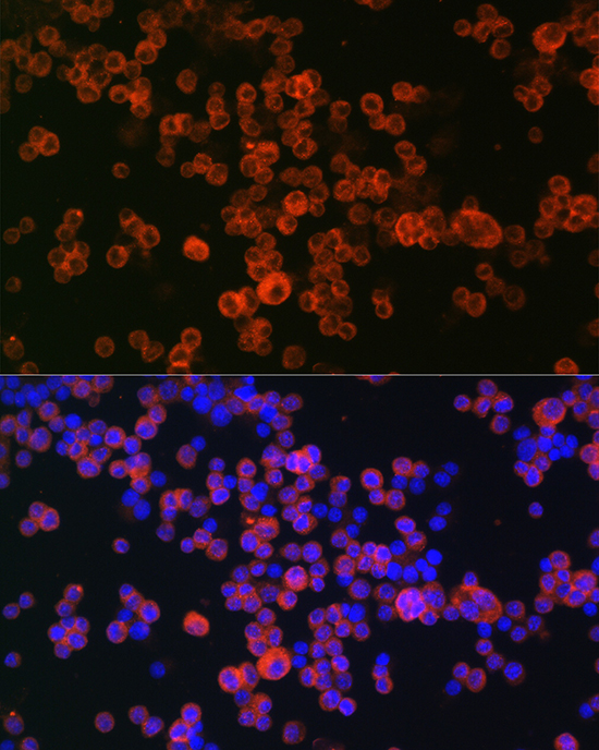
Immunofluorescence analysis of RAW264.7 cells using CD68 Rabbit pAb (A13286) at dilution of 1:50 (40x lens). Secondary antibody: Cy3-conjugated Goat anti-Rabbit IgG (H+L) (AS007) at 1:500 dilution. Blue: DAPI for nuclear staining. |
|
|
Immunofluorescence analysis of RAW264.7 cells using CD68 Rabbit pAb (A13286) at dilution of 1:50 (40x lens). Secondary antibody: Cy3-conjugated Goat anti-Rabbit IgG (H+L) (AS007) at 1:500 dilution. Blue: DAPI for nuclear staining. |
Product Guarantee and Expert Support
