
HNRNPD Rabbit pAb, Unconjugated, Polyclonal
Catalog Number:
ABB-A15679
- Images (9)


| Article Name: | HNRNPD Rabbit pAb, Unconjugated, Polyclonal |
| Biozol Catalog Number: | ABB-A15679 |
| Supplier Catalog Number: | A15679 |
| Alternative Catalog Number: | ABB-A15679-20UL,ABB-A15679-100UL,ABB-A15679-1000UL,ABB-A15679-500UL |
| Manufacturer: | ABclonal |
| Host: | Rabbit |
| Category: | Antikörper |
| Application: | ELISA, IF, IHC-P, IP, WB |
| Species Reactivity: | Human |
| Immunogen: | Recombinant protein (or fragment).This information is considered to be commercially sensitive. |
| Conjugation: | Unconjugated |
| Alternative Names: | P37, AUF1, AUF1A, HNRPD, hnRNPD0, HNRNPD |
| This gene belongs to the subfamily of ubiquitously expressed heterogeneous nuclear ribonucleoproteins (hnRNPs). The hnRNPs are nucleic acid binding proteins and they complex with heterogeneous nuclear RNA (hnRNA). These proteins are associated with pre-mRNAs in the nucleus and appear to influence pre-mRNA processing and other aspects of mRNA metabolism and transport. While all of the hnRNPs are present in the nucleus, some seem to shuttle between the nucleus and the cytoplasm. The hnRNP proteins have distinct nucleic acid binding properties. The protein encoded by this gene has two repeats of quasi-RRM domains that bind to RNAs. It localizes to both the nucleus and the cytoplasm. This protein is implicated in the regulation of mRNA stability. Alternative splicing of this gene results in four transcript variants. |
| Clonality: | Polyclonal |
| Molecular Weight: | 38kDa |
| NCBI: | 3184 |
| UniProt: | Q14103 |
| Purity: | Affinity purification |
| Sequence: | MSEEQFGGDGAAAAATAAVGGSAGEQEGAMVAATQGAAAAAGSGAGTGGGTASGGTEGGSAESEGAKIDASKNEEDEGHSNSSPRHSEAATAQREEWKMFIGGLSWDTTKKDLKDYFSKFGEVVDCTLKLDPITGRSRGFGFVLFKESESVDKVMDQKEHKLNGKVIDPKRAKAMKTKEPVKKIFVGGLSPDTPEEKIREYFGGFGEVESIELPMDNKTNKRRGFCFITFKEEEPVKKIMEKKYHNVGLSKCEIK |
| Target: | HNRNPD |
| Antibody Type: | Primary Antibody |
| Application Dilute: | WB,1:500 - 1:2000|IHC-P,1:50 - 1:200|IF/ICC,1:50 - 1:200|IP,0.5µg-4µg antibody for 200µg-400µg extracts of whole cells|ELISA,Recommended starting concentration is 1 µg/mL. Please optimize the concentration based on your specific assay requirements. |
| Application Notes: | Cross-Reactivity: Human,Mouse,Rat. ResearchArea: Epigenetics Nuclear Signaling,RNA Binding. Shipping: Ice Bag |
|
|
Immunohistochemistry analysis of paraffin-embedded Rat liver using HNRNPD Rabbit pAb (A15679) at dilution of 1:100 (40x lens). Microwave antigen retrieval performed with 0.01M PBS Buffer (pH 7.2) prior to IHC staining. |
|
|
Immunohistochemistry analysis of paraffin-embedded Human lung cancer using HNRNPD Rabbit pAb (A15679) at dilution of 1:100 (40x lens). Microwave antigen retrieval performed with 0.01M PBS Buffer (pH 7.2) prior to IHC staining. |
|
|
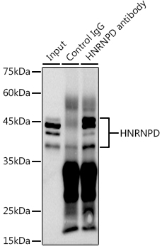
Western blot analysis of various lysates using HNRNPD Rabbit pAb (A15679) at 1:1000 dilution. Secondary antibody: HRP-conjugated Goat anti-Rabbit IgG (H+L) (AS014) at 1:10000 dilution. Lysates/proteins: 25µg per lane. Blocking buffer: 3% nonfat dry milk in TBST. Detection: ECL Basic Kit (RM00020). Exposure time: 30s. |
|
|
Immunohistochemistry analysis of paraffin-embedded Mouse brain using HNRNPD Rabbit pAb (A15679) at dilution of 1:100 (40x lens). Microwave antigen retrieval performed with 0.01M PBS Buffer (pH 7.2) prior to IHC staining. |
|
|
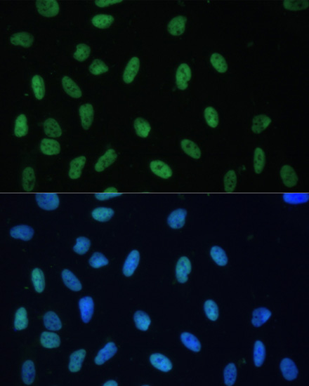
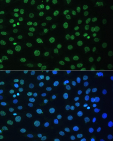
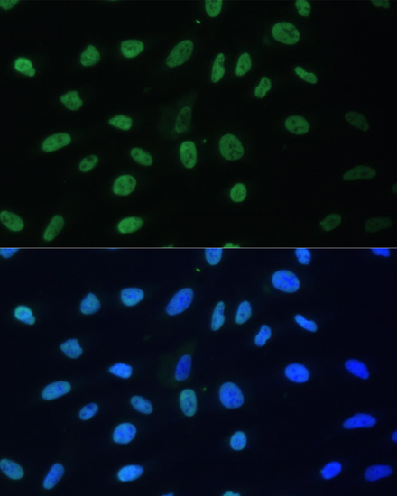
Immunofluorescence analysis of C6 cells using HNRNPD Rabbit pAb (A15679) at dilution of 1:100. Secondary antibody: Cy3-conjugated Goat anti-Rabbit IgG (H+L) (AS007) at 1:500 dilution. Blue: DAPI for nuclear staining. |
|
|
Immunofluorescence analysis of U-2 OS cells using HNRNPD Rabbit pAb (A15679) at dilution of 1:100. Secondary antibody: Cy3-conjugated Goat anti-Rabbit IgG (H+L) (AS007) at 1:500 dilution. Blue: DAPI for nuclear staining. |
|
|
Immunoprecipitation analysis of 300 µg extracts of HeLa cells using 3 µg HNRNPD antibody (A15679). Western blot was performed from the immunoprecipitate using HNRNPD antibody (A15679) at a dilution of 1:1000. |
Product Guarantee and Expert Support
