
CD11b/ITGAM Rabbit pAb, Unconjugated, Polyclonal
Catalog Number:
ABB-A1581
- Images (9)


| Article Name: | CD11b/ITGAM Rabbit pAb, Unconjugated, Polyclonal |
| Biozol Catalog Number: | ABB-A1581 |
| Supplier Catalog Number: | A1581 |
| Alternative Catalog Number: | ABB-A1581-100UL,ABB-A1581-20UL,ABB-A1581-1000UL,ABB-A1581-500UL |
| Manufacturer: | ABclonal |
| Host: | Rabbit |
| Category: | Antikörper |
| Application: | ELISA, IF, IHC-P, WB |
| Species Reactivity: | Human |
| Immunogen: | Recombinant protein (or fragment).This information is considered to be commercially sensitive. |
| Conjugation: | Unconjugated |
| Alternative Names: | CR3A, MO1A, CD11B, MAC-1, MAC1A, SLEB6, CD11b/ITGAM |
| This gene encodes the integrin alpha M chain. Integrins are heterodimeric integral membrane proteins composed of an alpha chain and a beta chain. This I-domain containing alpha integrin combines with the beta 2 chain (ITGB2) to form a leukocyte-specific integrin referred to as macrophage receptor 1 (Mac-1), or inactivated-C3b (iC3b) receptor 3 (CR3). The alpha M beta 2 integrin is important in the adherence of neutrophils and monocytes to stimulated endothelium, and also in the phagocytosis of complement coated particles. Multiple transcript variants encoding different isoforms have been found for this gene. |
| Clonality: | Polyclonal |
| Molecular Weight: | 127kDa |
| NCBI: | 3684 |
| UniProt: | P11215 |
| Purity: | Affinity purification |
| Sequence: | PQEDSDIAFLIDGSGSIIPHDFRRMKEFVSTVMEQLKKSKTLFSLMQYSEEFRIHFTFKEFQNNPNPRSLVKPITQLLGRTHTATGIRKVVRELFNITNGARKNAFKILVVITDGEKFGDPLGYEDVIPEADREGVIRYVIGVGDAFRSEKSRQELNTIASKPPRDHVFQVNNFEALKTIQNQLREKIFAIEGTQT |
| Target: | ITGAM |
| Antibody Type: | Primary Antibody |
| Application Dilute: | WB,1:100 - 1:500|IHC-P,1:50 - 1:200|IF/ICC,1:50 - 1:200|ELISA,Recommended starting concentration is 1 µg/mL. Please optimize the concentration based on your specific assay requirements. |
| Application Notes: | Cross-Reactivity: Human,Mouse,Rat. ResearchArea: Signal Transduction,G protein signaling,G-Protein-Coupled Receptors Signaling to MAPK Erk,PI3K-Akt Signaling Pathway,MAPK-Erk Signaling Pathway,Cell Biology Developmental Biology,Cell Adhesion,Cytoskeleton,Immunology Inflammation,CDs,Neuroscience, Cell Type Marker,Stem Cells,Hematopoietic Progenitors,Mesenchymal Stem Cells. Shipping: Ice Bag |
|
|
Immunohistochemistry analysis of paraffin-embedded Human tonsil using CD11b/ITGAM Rabbit pAb (A1581) at dilution of 1:100 (40x lens). High pressure antigen retrieval performed with 0.01M Citrate buffer (pH 6.0) prior to IHC staining. |
|
|
Immunohistochemistry analysis of paraffin-embedded Mouse spleen using CD11b/ITGAM Rabbit pAb (A1581) at dilution of 1:100 (40x lens). High pressure antigen retrieval performed with 0.01M Citrate buffer (pH 6.0) prior to IHC staining. |
|
|
Western blot analysis of various lysates using CD11b/ITGAM Rabbit pAb (A1581) at 1:500 dilution. Secondary antibody: HRP-conjugated Goat anti-Rabbit IgG (H+L) (AS014) at 1:10000 dilution. Lysates/proteins: 25µg per lane. Blocking buffer: 3% nonfat dry milk in TBST. Detection: ECL Enhanced Kit (RM00021). Exposure time: 90s. |
|
|
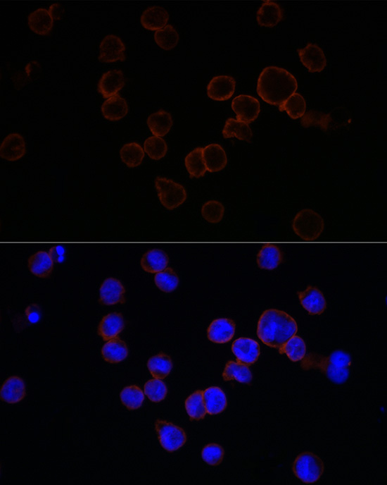
Immunofluorescence analysis of TF-1 cells using CD11b/ITGAM Rabbit pAb (A1581) at dilution of 1:100 (40x lens). Secondary antibody: Cy3-conjugated Goat anti-Rabbit IgG (H+L) (AS007) at 1:500 dilution. Blue: DAPI for nuclear staining. |
|
|
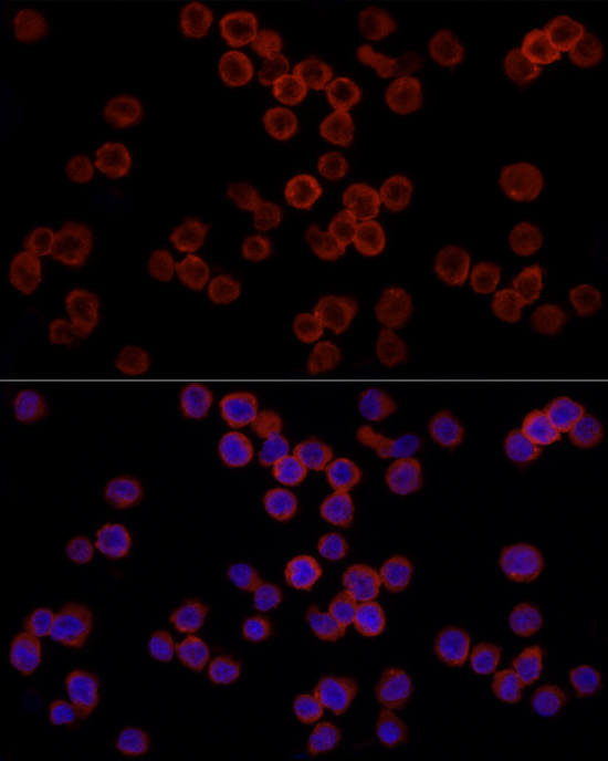
Immunofluorescence analysis of THP-1 cells using CD11b/ITGAM Rabbit pAb (A1581) at dilution of 1:100 (40x lens). Secondary antibody: Cy3-conjugated Goat anti-Rabbit IgG (H+L) (AS007) at 1:500 dilution. Blue: DAPI for nuclear staining. |
|
|
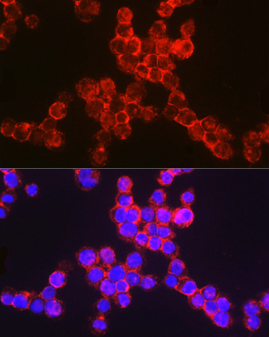
Immunofluorescence analysis of TF-1 cells using CD11b/ITGAM Rabbit pAb (A1581) at dilution of 1:100 (40x lens). Secondary antibody: Cy3-conjugated Goat anti-Rabbit IgG (H+L) (AS007) at 1:500 dilution. Blue: DAPI for nuclear staining. |
|
|
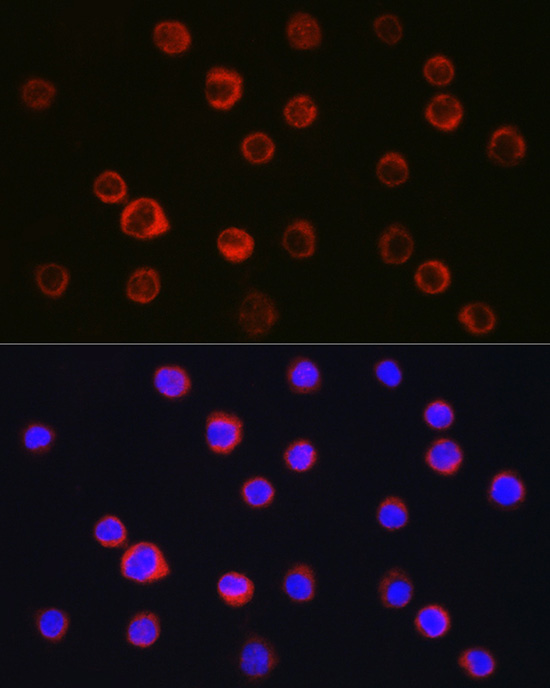
Immunofluorescence analysis of THP-1 cells using CD11b/ITGAM Rabbit pAb (A1581) at dilution of 1:100 (40x lens). Secondary antibody: Cy3-conjugated Goat anti-Rabbit IgG (H+L) (AS007) at 1:500 dilution. Blue: DAPI for nuclear staining. |
|
|
Immunofluorescence analysis of TF-1 cells using CD11b/ITGAM Rabbit pAb (A1581) at dilution of 1:100 (40x lens). Secondary antibody: Cy3-conjugated Goat anti-Rabbit IgG (H+L) (AS007) at 1:500 dilution. Blue: DAPI for nuclear staining. |
|
|
Immunofluorescence analysis of THP-1 cells using CD11b/ITGAM Rabbit pAb (A1581) at dilution of 1:100 (40x lens). Secondary antibody: Cy3-conjugated Goat anti-Rabbit IgG (H+L) (AS007) at 1:500 dilution. Blue: DAPI for nuclear staining. |
Product Guarantee and Expert Support
