
CSK Rabbit pAb, Polyclonal
Catalog Number:
ABB-A16824
- Images (9)
| Article Name: | CSK Rabbit pAb, Polyclonal |
| Biozol Catalog Number: | ABB-A16824 |
| Supplier Catalog Number: | A16824 |
| Alternative Catalog Number: | ABB-A16824-100UL,ABB-A16824-20UL,ABB-A16824-500UL,ABB-A16824-1000UL |
| Manufacturer: | ABclonal |
| Host: | Rabbit |
| Category: | Antikörper |
| Application: | ELISA, IF, IHC-P, WB |
| Species Reactivity: | Human |
| Immunogen: | Recombinant protein (or fragment).This information is considered to be commercially sensitive. |
| Alternative Names: | CSK |
| The protein encoded by this gene is involved in multiple pathways, including the regulation of Src family kinases. It plays an important role in T-cell activation through its association with the protein encoded by the protein tyrosine phosphatase, non-receptor type 22 (PTPN22) gene. This protein also phosphorylates C-terminal tyrosine residues on multiple substrates, including the protein encoded by the SRC proto-oncogene, non-receptor tyrosine kinase gene. Phosphorylation suppresses the kinase activity of the Src family tyrosine kinases. An intronic polymorphism (rs34933034) in this gene has been found to affect B-cell activation and is associated with systemic lupus erythematosus (SLE). Alternative splicing results in multiple transcript variants. |
| Clonality: | Polyclonal |
| Molecular Weight: | 51kDa |
| NCBI: | 1445 |
| UniProt: | P41240 |
| Purity: | Affinity purification |
| Sequence: | WALNMKELKLLQTIGKGEFGDVMLGDYRGNKVAVKCIKNDATAQAFLAEASVMTQLRHSNLVQLLGVIVEEKGGLYIVTEYMAKGSLVDYLRSRGRSVLGGDCLLKFSLDVCEAMEYLEGNNFVHRDLAARNVLVSEDNVAKVSDFGLTKEASSTQDTGKLPVKWTAPEALREKKFSTKSDVWSFGILLWEIYSFGRVPYPRIPLKDVVPRVEKGYKMDAPDGCPPAVYEVMKNCWHLDAAMRPSFLQLREQLEH |
| Target: | CSK |
| Antibody Type: | Primary Antibody |
| Application Dilute: | WB,1:500 - 1:1000|IHC-P,1:50 - 1:200|IF/ICC,1:50 - 1:200|ELISA,Recommended starting concentration is 1 µg/mL. Please optimize the concentration based on your specific assay requirements. |
| Application Notes: | Cross-Reactivity: Human,Mouse,Rat. ResearchArea: Signal Transduction,Kinase,Tyrosine kinases,Immunology Inflammation,B Cell Receptor Signaling Pathway. Shipping: Ice Bag |
|
|
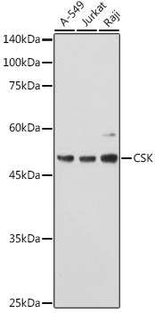
Western blot analysis of various lysates using CSK Rabbit pAb (A16824) at 1:1000 dilution. Secondary antibody: HRP-conjugated Goat anti-Rabbit IgG (H+L) (AS014) at 1:10000 dilution. Lysates/proteins: 25µg per lane. Blocking buffer: 3% nonfat dry milk in TBST. Detection: ECL Basic Kit (RM00020). Exposure time: 60s. |
|
|
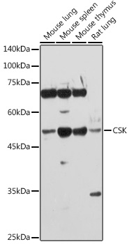
Western blot analysis of various lysates using CSK Rabbit pAb (A16824) at 1:1000 dilution. Secondary antibody: HRP-conjugated Goat anti-Rabbit IgG (H+L) (AS014) at 1:10000 dilution. Lysates/proteins: 25µg per lane. Blocking buffer: 3% nonfat dry milk in TBST. Detection: ECL Basic Kit (RM00020). Exposure time: 90s. |
|
|
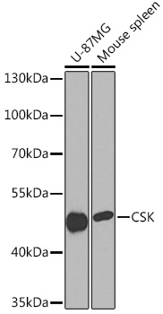
Immunohistochemistry analysis of paraffin-embedded Rat spleen using CSK Rabbit pAb (A16824) at dilution of 1:100 (40x lens). High pressure antigen retrieval performed with 0.01M Citrate buffer (pH 6.0) prior to IHC staining. |
|
|
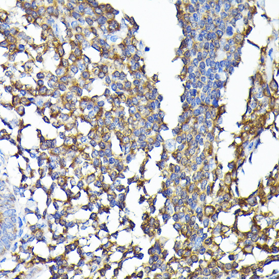
Immunohistochemistry analysis of paraffin-embedded Human colon carcinoma using CSK Rabbit pAb (A16824) at dilution of 1:100 (40x lens). High pressure antigen retrieval performed with 0.01M Citrate buffer (pH 6.0) prior to IHC staining. |
|
|
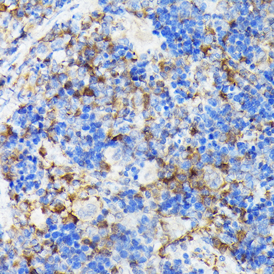
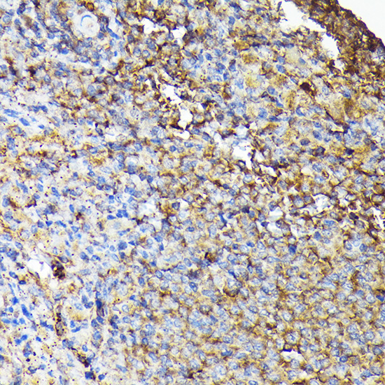
Immunohistochemistry analysis of paraffin-embedded Mouse spleen using CSK Rabbit pAb (A16824) at dilution of 1:100 (40x lens). High pressure antigen retrieval performed with 0.01M Citrate buffer (pH 6.0) prior to IHC staining. |
|
|
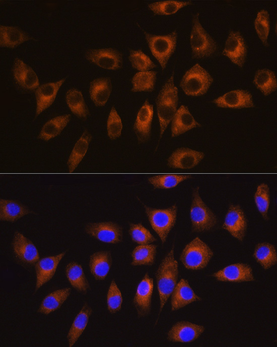
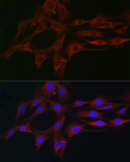
Immunofluorescence analysis of L929 cells using CSK Rabbit pAb (A16824) at dilution of 1:100 (40x lens). Secondary antibody: Cy3-conjugated Goat anti-Rabbit IgG (H+L) (AS007) at 1:500 dilution. Blue: DAPI for nuclear staining. |
|
|
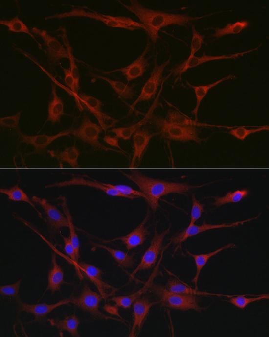
Immunofluorescence analysis of C6 cells using CSK Rabbit pAb (A16824) at dilution of 1:100 (40x lens). Secondary antibody: Cy3-conjugated Goat anti-Rabbit IgG (H+L) (AS007) at 1:500 dilution. Blue: DAPI for nuclear staining. |
|
|
Immunofluorescence analysis of NIH/3T3 cells using CSK Rabbit pAb (A16824) at dilution of 1:100 (40x lens). Secondary antibody: Cy3-conjugated Goat anti-Rabbit IgG (H+L) (AS007) at 1:500 dilution. Blue: DAPI for nuclear staining. |
Product Guarantee and Expert Support
